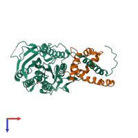
3h4s

X-ray diffraction
2.4Å resolution

Structure of the complex of a mitotic kinesin with its calcium binding regulator

Released:
Source organism: Arabidopsis thaliana
Primary publication:
Structure of the complex of a mitotic kinesin with its calcium binding regulator.
Proc Natl Acad Sci U S A 106 8175-9 (2009)
PMID: 19416847

Function and Biology Details

Structure analysis Details

Assembly composition:
hetero dimer (preferred)
PDBe Complex ID:
PDB-CPX-190318 (preferred)
Entry contents:
2 distinct polypeptide molecules
Macromolecules (2 distinct):
Kinesin-like protein KIN-14E Chain: A
Molecule details ›
Chain: A
Length: 386 amino acids
Theoretical weight: 44.04 KDa
Source organism: Arabidopsis thaliana
Expression system: Escherichia coli
UniProt:
  • Canonical: Q9FHN8 (Residues: 875-1260; Coverage: 31%)
Gene names: At5g65930, K14B20.10, KCBP, KIN14E, ZWI
Sequence domains: Kinesin motor domain
Structure domains:
Calcium-binding protein KIC Chain: E
Molecule details ›
Chain: E
Length: 135 amino acids
Theoretical weight: 15.18 KDa
Source organism: Arabidopsis thaliana
Expression system: Escherichia coli
UniProt:
  • Canonical: Q9ZPX9 (Residues: 1-135; Coverage: 100%)
Gene names: At2g46600, F13A10.13, KIC
Structure domains: EF-hand

Ligands and Environments

3 bound ligands:
No modified residues

Experiments and Validation Details

Entry percentile scores
X-ray source: ALS BEAMLINE 8.3.1
Spacegroup: P6522
Unit cell:
a: 118.8Å b: 118.8Å c: 142.1Å
α: 90° β: 90° γ: 120°
R-values:
R R work R free
0.223 0.223 0.228
Expression system: Escherichia coli