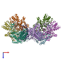
3exh

X-ray diffraction
2.44Å resolution

Crystal structure of the pyruvate dehydrogenase (E1p) component of human pyruvate dehydrogenase complex

Released:

Function and Biology Details

Structure analysis Details

Assembly composition:
hetero tetramer (preferred)
PDBe Complex ID:
PDB-CPX-140173 (preferred)
Entry contents:
3 distinct polypeptide molecules
Macromolecules (3 distinct):
Pyruvate dehydrogenase E1 component subunit alpha, somatic form, mitochondrial Chains: A, E
Molecule details ›
Chains: A, E
Length: 382 amino acids
Theoretical weight: 42.55 KDa
Source organism: Homo sapiens
Expression system: Escherichia coli
UniProt:
  • Canonical: P08559 (Residues: 30-390; Coverage: 93%)
Gene names: PDHA1, PHE1A
Sequence domains: Dehydrogenase E1 component
Structure domains: Rossmann fold
Pyruvate dehydrogenase E1 component subunit beta, mitochondrial Chains: B, D, F, H
Molecule details ›
Chains: B, D, F, H
Length: 329 amino acids
Theoretical weight: 35.94 KDa
Source organism: Homo sapiens
Expression system: Escherichia coli
UniProt:
  • Canonical: P11177 (Residues: 31-359; Coverage: 92%)
Gene names: PDHB, PHE1B
Sequence domains:
Structure domains:
Pyruvate dehydrogenase E1 component subunit alpha, somatic form, mitochondrial Chains: C, G
Molecule details ›
Chains: C, G
Length: 382 amino acids
Theoretical weight: 42.63 KDa
Source organism: Homo sapiens
Expression system: Escherichia coli
UniProt:
  • Canonical: P08559 (Residues: 30-390; Coverage: 93%)
Gene names: PDHA1, PHE1A
Sequence domains: Dehydrogenase E1 component
Structure domains: Rossmann fold

Ligands and Environments


Cofactor: Ligand TPP 4 x TPP
3 bound ligands:
1 modified residue:

Experiments and Validation Details

Entry percentile scores
X-ray source: RIGAKU FR-D
Spacegroup: C2
Unit cell:
a: 257.064Å b: 115.612Å c: 127.592Å
α: 90° β: 113.64° γ: 90°
R-values:
R R work R free
0.167 0.165 0.211
Expression system: Escherichia coli