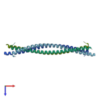
3bat

X-ray diffraction
2.3Å resolution

Crystal structure of the N-terminal region of the scallop myosin rod, monoclinic (P21) form

Released:

Function and Biology Details

Biochemical function:
  • not assigned
Biological process:
  • not assigned
Cellular component:
  • not assigned

Structure analysis Details

Assembly composition:
homo dimer (preferred)
PDBe Complex ID:
PDB-CPX-136510 (preferred)
Entry contents:
1 distinct polypeptide molecule
Macromolecule:
General control transcription factor GCN4; Myosin heavy chain, striated muscle Chains: A, B, C, D
Molecule details ›
Chains: A, B, C, D
Length: 89 amino acids
Theoretical weight: 10.56 KDa
Source organisms: Expression system: Escherichia coli
UniProt:
  • Canonical: P24733 (Residues: 835-885; Coverage: 3%)
  • Canonical: P03069 (Residues: 250-281; Coverage: 11%)
Gene names: AAS101, AAS3, ARG9, GCN4, YEL009C
Sequence domains: Basic region leucine zipper
Structure domains: Single alpha-helices involved in coiled-coils or other helix-helix interfaces

Ligands and Environments

No bound ligands
No modified residues

Experiments and Validation Details

Entry percentile scores
X-ray source: NSLS BEAMLINE X26C
Spacegroup: P21
Unit cell:
a: 65.782Å b: 40.217Å c: 77.404Å
α: 90° β: 111.54° γ: 90°
R-values:
R R work R free
0.253 0.253 0.299
Expression system: Escherichia coli