2q8o

X-ray diffraction
1.75Å resolution

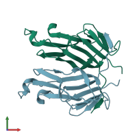
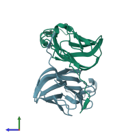
crystal structure of mouse GITR ligand dimer

Released:
Source organism: Mus musculus
Primary publication:
Structural basis for ligand-mediated mouse GITR activation.
Proc Natl Acad Sci U S A 105 641-5 (2008)
PMID: 18178614

Function and Biology Details

Structure analysis Details

Assembly composition:
homo dimer (preferred)
PDBe Complex ID:
PDB-CPX-182023 (preferred)
Entry contents:
1 distinct polypeptide molecule
Macromolecule:
Tumor necrosis factor ligand superfamily member 18 Chains: A, B
Molecule details ›
Chains: A, B
Length: 136 amino acids
Theoretical weight: 15.54 KDa
Source organism: Mus musculus
Expression system: Escherichia coli BL21
UniProt:
  • Canonical: Q7TS55 (Residues: 43-173; Coverage: 76%)
Gene names: Gitrl, Tnfsf18
Sequence domains: TNF(Tumour Necrosis Factor) family
Structure domains: Jelly Rolls

Ligands and Environments

No bound ligands
No modified residues

Experiments and Validation Details

Entry percentile scores
X-ray source: RIGAKU MICROMAX-007
Spacegroup: P212121
Unit cell:
a: 52.556Å b: 70.402Å c: 71.647Å
α: 90° β: 90° γ: 90°
R-values:
R R work R free
0.18 0.18 0.221
Expression system: Escherichia coli BL21