2i3t

X-ray diffraction
2.8Å resolution

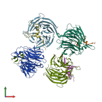
Bub3 complex with Mad3 (BubR1) GLEBS motif

Released:
Source organism: Saccharomyces cerevisiae
Primary publication:
Structural analysis of Bub3 interactions in the mitotic spindle checkpoint.
Proc Natl Acad Sci U S A 104 1201-6 (2007)
PMID: 17227844

Function and Biology Details

Structure analysis Details

Assembly composition:
hetero dimer (preferred)
PDBe Complex ID:
PDB-CPX-150755 (preferred)
Entry contents:
2 distinct polypeptide molecules
Macromolecules (2 distinct):
Cell cycle arrest protein BUB3 Chains: A, C, E, G
Molecule details ›
Chains: A, C, E, G
Length: 341 amino acids
Theoretical weight: 38.48 KDa
Source organism: Saccharomyces cerevisiae
Expression system: Escherichia coli
UniProt:
  • Canonical: P26449 (Residues: 1-341; Coverage: 100%)
Gene names: BUB3, OR26.16, YOR026W
Sequence domains: WD domain, G-beta repeat
Structure domains: YVTN repeat-like/Quinoprotein amine dehydrogenase
Spindle assembly checkpoint component MAD3 Chains: B, D, F, H
Molecule details ›
Chains: B, D, F, H
Length: 54 amino acids
Theoretical weight: 6.58 KDa
Source organism: Saccharomyces cerevisiae
Expression system: Escherichia coli
UniProt:
  • Canonical: P47074 (Residues: 353-400; Coverage: 9%)
Gene names: J1341, MAD3, YJL013C
Sequence domains: Mad3/BUB1 homology region 2
Structure domains: N-terminal domain of TfIIb

Ligands and Environments

No bound ligands
No modified residues

Experiments and Validation Details

Entry percentile scores
X-ray source: ALS BEAMLINE 8.2.2
Spacegroup: P21
Unit cell:
a: 51.352Å b: 173.335Å c: 90.019Å
α: 90° β: 95.3° γ: 90°
R-values:
R R work R free
0.242 0.238 0.267
Expression system: Escherichia coli