Function and Biology Details
Reaction catalysed:
Methylamine + H(2)O + 2 oxidized [amicyanin] = formaldehyde + NH(3) + 2 reduced [amicyanin]
Biochemical function:
Biological process:
Cellular component:
Sequence domains:
- Methylamine/Aralkylamine dehydrogenase light chain superfamily
- Methylamine/Aralkylamine dehydrogenase light chain, C-terminal domain
- Methylamine dehydrogenase light chain
- Amine dehydrogenase heavy chain
- Quinoprotein amine dehydrogenase, beta chain-like
- WD40/YVTN repeat-like-containing domain superfamily
- Methylamine dehydrogenase heavy chain
Structure domains:
Structure analysis Details
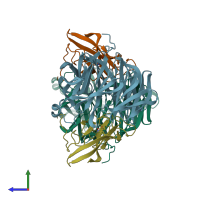
Assembly composition:
hetero tetramer (preferred)
Assembly name:
Methylamine dehydrogenase (preferred)
PDBe Complex ID:
PDB-CPX-149679 (preferred)
Entry contents:
2 distinct polypeptide molecules
Macromolecules (2 distinct):


