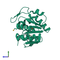
1pdv

X-ray diffraction
1.8Å resolution

Crystal structure of human DJ-1, P 31 2 1 space group

Released:
Source organism: Homo sapiens
Primary publication:
Crystal structure of human DJ-1, a protein associated with early onset Parkinson's disease.
J Biol Chem 278 31372-9 (2003)
PMID: 12761214

Function and Biology Details

Reaction catalysed:
An N(6)-(1-hydroxy-2-oxopropyl)-[protein]-L-lysine + H(2)O = a [protein]-L-lysine + lactate

Structure analysis Details

Assembly composition:
homo dimer (preferred)
PDBe Complex ID:
PDB-CPX-189207 (preferred)
Entry contents:
1 distinct polypeptide molecule
Macromolecule:
Parkinson disease protein 7 Chain: A
Molecule details ›
Chain: A
Length: 197 amino acids
Theoretical weight: 21.18 KDa
Source organism: Homo sapiens
Expression system: Escherichia coli
UniProt:
  • Canonical: Q99497 (Residues: 1-189; Coverage: 100%)
Gene name: PARK7
Sequence domains: DJ-1/PfpI family
Structure domains: Rossmann fold

Ligands and Environments

No bound ligands
1 modified residue:

Experiments and Validation Details

Entry percentile scores
X-ray source: NSLS BEAMLINE X4A
Spacegroup: P3121
Unit cell:
a: 75.658Å b: 75.658Å c: 75.488Å
α: 90° β: 90° γ: 120°
R-values:
R R work R free
0.19 0.19 0.215
Expression system: Escherichia coli