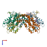
1lia

X-ray diffraction
2.8Å resolution

CRYSTAL STRUCTURE OF R-PHYCOERYTHRIN FROM POLYSIPHONIA AT 2.8 A RESOLUTION

Released:

Function and Biology Details

Biochemical function:
  • not assigned
Biological process:
Cellular component:

Structure analysis Details

Assemblies composition:
hetero dimer (preferred)
hetero dodecamer
hetero hexamer
hetero tetramer
PDBe Complex ID:
PDB-CPX-160920 (preferred)
Entry contents:
2 distinct polypeptide molecules
Macromolecules (2 distinct):
R-phycoerythrin alpha chain Chains: A, K
Molecule details ›
Chains: A, K
Length: 164 amino acids
Theoretical weight: 17.86 KDa
Source organism: Polysiphonia urceolata
UniProt:
  • Canonical: P84861 (Residues: 1-164; Coverage: 100%)
Gene names: cpeA, rpeA
Sequence domains: Phycobilisome protein
Structure domains: Phycocyanins
R-phycoerythrin beta chain Chains: B, L
Molecule details ›
Chains: B, L
Length: 177 amino acids
Theoretical weight: 18.74 KDa
Source organism: Polysiphonia urceolata
UniProt:
  • Canonical: P84862 (Residues: 1-177; Coverage: 100%)
Gene names: cpeB, rpeB
Sequence domains: Phycobilisome protein
Structure domains: Phycocyanins

Ligands and Environments

2 bound ligands:
No modified residues

Experiments and Validation Details

Entry percentile scores
Spacegroup: R3
Unit cell:
a: 189.8Å b: 189.8Å c: 60.1Å
α: 90° β: 90° γ: 120°
R-values:
R R work R free
0.18 0.18 0.182