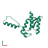
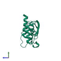
1k46

X-ray diffraction
2.2Å resolution

Crystal Structure of the Type III Secretory Domain of Yersinia YopH Reveals a Domain-Swapped Dimer

Released:

Function and Biology Details

Reaction catalysed:
Protein tyrosine phosphate + H(2)O = protein tyrosine + phosphate
Biological process:
Cellular component:
  • not assigned

Structure analysis Details

Assemblies composition:
monomeric (preferred)
homo dimer
PDBe Complex ID:
PDB-CPX-140166 (preferred)
Entry contents:
1 distinct polypeptide molecule
Macromolecule:
Tyrosine-protein phosphatase YopH Chain: A
Molecule details ›
Chain: A
Length: 136 amino acids
Theoretical weight: 14.88 KDa
Source organism: Yersinia pseudotuberculosis
Expression system: Escherichia coli BL21(DE3)
UniProt:
  • Canonical: P08538 (Residues: 1-129; Coverage: 28%)
Gene names: pYV0094, yop2b, yopH
Sequence domains: YopH, N-terminal
Structure domains: Protein-tyrosine phosphatase, YopH, N-terminal domain

Ligands and Environments

No bound ligands
No modified residues

Experiments and Validation Details

Entry percentile scores
X-ray source: RIGAKU RUH3R
Spacegroup: C2221
Unit cell:
a: 47.9Å b: 120.7Å c: 48.8Å
α: 90° β: 90° γ: 90°
R-values:
R R work R free
0.224 0.224 0.257
Expression system: Escherichia coli BL21(DE3)