1bvp

X-ray diffraction
2.6Å resolution

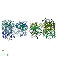
THE CRYSTAL STRUCTURE OF BLUETONGUE VIRUS VP7

Released:
Primary publication:
The crystal structure of bluetongue virus VP7.
Nature 373 167-70 (1995)
PMID: 7816101

Function and Biology Details

Structure analysis Details

Assemblies composition:
homo trimer (preferred)
homo hexamer
Assembly name:
PDBe Complex ID:
PDB-CPX-159577 (preferred)
Entry contents:
1 distinct polypeptide molecule
Macromolecule:
Core protein VP7 Chains: 1, 2, 3, 4, 5, 6
Molecule details ›
Chains: 1, 2, 3, 4, 5, 6
Length: 349 amino acids
Theoretical weight: 38.58 KDa
Source organism: Bluetongue virus (serotype 10 / American isolate)
Expression system: Not provided
UniProt:
  • Canonical: P69361 (Residues: 1-349; Coverage: 100%)
Gene name: Segment-7
Sequence domains: Orbivirus inner capsid protein VP7
Structure domains:

Ligands and Environments

No bound ligands
No modified residues

Experiments and Validation Details

Entry percentile scores
Spacegroup: P21
Unit cell:
a: 83.6Å b: 110.2Å c: 129.8Å
α: 90° β: 103.1° γ: 90°
R-values:
R R work R free
0.184 0.184 not available
Expression system: Not provided