
|
|
|
|
|
 |
Contents |
 |
|
|
|
|
|
|
|
|
|
|
|

 430 a.a.
|
|
|
|
|
|
|
|
|
|
|

 352 a.a.
|
|
|
|
|
|
|
|
|
|
|

 385 a.a.
|
|
|
|
|
|
|
|
|
|
|

 245 a.a.
|
|
|
|
|
|
|
|
|
|
|

 185 a.a.
|
|
|
|
|
|
|
|
|
|
|

 74 a.a.
|
|
|
|
|
|
|
|
|
|
|

 125 a.a.
|
|
|
|
|
|
|
|
|
|
|

 93 a.a.
|
|
|
|
|
|
|
|
|
|
|

 53 a.a.
|
|
|
|
|
|
|
|
|
|
|

 127 a.a.
|
|
|
|
|
|
|
|
|
|
|

 107 a.a.
|
|
|
|
|
|
|
|
|
|
|
 108 a.a.
|
|
|
|
|
|
|
|
|
* Residue conservation analysis
|
|
|
|
|
|
PDB id:
|
|
|
|
| Name: |
|
Oxidoreductase/electron transport
|
|
|
Title:
|
|
Yeast cytochrome bc1 complex with bound substrate cytochromE C
|
|
Structure:
|
|
Ubiquinol-cytochromE C reductase complex core protein i. Chain: a, l. Fragment: residues 27-457. Ubiquinol-cytochromE C reductase complex core protein 2. Chain: b, m. Fragment: residues 17-368. Cytochrome b. Chain: c, n. Cytochrome c1, heme protein.
|
|
Source:
|
|
Saccharomyces cerevisiae. Baker's yeast. Organism_taxid: 4932. Organelle: mitochondria. Mus musculus. House mouse. Organism_taxid: 10090. Expressed in: escherichia coli. Expression_system_taxid: 562.
|
|
Biol. unit:
|
|
23mer (from
)
|
|
Resolution:
|
|
|
2.97Å
|
R-factor:
|
0.229
|
R-free:
|
0.268
|
|
|
Authors:
|
|
C.Lange,C.Hunte
|
Key ref:

|
|
C.Lange
and
C.Hunte
(2002).
Crystal structure of the yeast cytochrome bc1 complex with its bound substrate cytochrome c.
Proc Natl Acad Sci U S A,
99,
2800-2805.
PubMed id:
DOI:

|
|
|
Date:
|
|
|
05-Feb-02
|
Release date:
|
06-Mar-02
|
|
|
|
|
|
|
PROCHECK
|

|
|
|
|
|
Headers
|
|
|
|
References
|
|
|
|
|
|

|
|
P07256
(QCR1_YEAST) -
Cytochrome b-c1 complex subunit 1, mitochondrial from Saccharomyces cerevisiae (strain ATCC 204508 / S288c)
|
|
|
|
Seq:
Struc:
|
|
|
|
457 a.a.
430 a.a.*
|
|

|

|

|
|
|
|
|
|
|
|
|
|
|
|

|
|
P07257
(QCR2_YEAST) -
Cytochrome b-c1 complex subunit 2, mitochondrial from Saccharomyces cerevisiae (strain ATCC 204508 / S288c)
|
|
|
|
Seq:
Struc:
|
|
|
|
368 a.a.
352 a.a.
|
|
|

|

|
|
|
|
|
|
|
|
|
|
|
|

|
|
P00163
(CYB_YEAST) -
Cytochrome b from Saccharomyces cerevisiae (strain ATCC 204508 / S288c)
|
|
|
|
Seq:
Struc:
|
|
|
|
385 a.a.
385 a.a.*
|
|
|

|

|
|
|
|
|
|
|
|
|
|
|
|

|
|
P07143
(CY1_YEAST) -
Cytochrome c1, heme protein, mitochondrial from Saccharomyces cerevisiae (strain ATCC 204508 / S288c)
|
|
|
|
Seq:
Struc:
|
|
|
|
309 a.a.
245 a.a.
|
|
|

|

|
|
|
|
|
|
|
|
|
|
|
|

|
|
P08067
(UCRI_YEAST) -
Cytochrome b-c1 complex subunit Rieske, mitochondrial from Saccharomyces cerevisiae (strain ATCC 204508 / S288c)
|
|
|
|
Seq:
Struc:
|
|
|
|
215 a.a.
185 a.a.
|
|
|

|
|
|
|
|
|
|
|
|
|
|
|
|

|
|
P00127
(QCR6_YEAST) -
Cytochrome b-c1 complex subunit 6, mitochondrial from Saccharomyces cerevisiae (strain ATCC 204508 / S288c)
|
|
|
|
Seq:
Struc:
|
|
|
|
147 a.a.
74 a.a.*
|
|
|

|
|
|
|
|
|
|
|
|
|
|
|
|

|
|
P00128
(QCR7_YEAST) -
Cytochrome b-c1 complex subunit 7, mitochondrial from Saccharomyces cerevisiae (strain ATCC 204508 / S288c)
|
|
|
|
Seq:
Struc:
|
|
|
|
127 a.a.
125 a.a.
|
|
|

|
|
|
|
|
|
|
|
|
|
|
|
|

|
|
P08525
(QCR8_YEAST) -
Cytochrome b-c1 complex subunit 8, mitochondrial from Saccharomyces cerevisiae (strain ATCC 204508 / S288c)
|
|
|
|
Seq:
Struc:
|
|
|
|
94 a.a.
93 a.a.
|
|
|

|
|
|
|
|
|
|
|
|
|
|
|
|

|
|
P22289
(QCR9_YEAST) -
Cytochrome b-c1 complex subunit 9, mitochondrial from Saccharomyces cerevisiae (strain ATCC 204508 / S288c)
|
|
|
|
Seq:
Struc:
|
|
|
|
66 a.a.
53 a.a.*
|
|
|

|
|
|
|
|
|
|
|
|
|
|
|
|

|
|
No UniProt id for this chain
|
|
|
|
|
|
|
|
|
|
|
|
|
|
|
|
|
|
|
|
|
Enzyme class 2:
|
|
Chains A, B, F, G, H, I, L, M, Q, R, S, T:
E.C.1.10.2.2
- Transferred entry: 7.1.1.8.
|
|
|
|
|
|
|
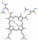
Reaction:
|
|
Quinol + 2 ferricytochrome c = quinone + 2 ferrocytochrome c + 2 H+
|
|
|
|
|
|
Quinol
|
+
|
 2
×
ferricytochrome c
2
×
ferricytochrome c
|
=
|
 quinone
|
+
|
 2
×
ferrocytochrome c
2
×
ferrocytochrome c
|
+
|
2
×
H(+)
|
|
|
|
|
|
|
|
|
|
Enzyme class 3:
|
|
Chains C, D, E, N, O, P:
E.C.7.1.1.8
- quinol--cytochrome-c reductase.
|
|
|
|
|
|
|
Reaction:
|
|
a quinol + 2 Fe(III)-[cytochrome c](out) = a quinone + 2 Fe(II)- [cytochrome c](out) + 2 H(+)(out)
|
|
|
|
|
|
quinol
|
+
|
2
×
Fe(III)-[cytochrome c](out)
|
=
|
quinone
|
+
|
2
×
Fe(II)- [cytochrome c](out)
|
+
|
2
×
H(+)(out)
|
|
|
|
|
|
|
|
|
|
Enzyme class 4:
|
|
Chain W:
E.C.?
|
|
|
|
|
|
|
|
|
|
Note, where more than one E.C. class is given (as above), each may
correspond to a different protein domain or, in the case of polyprotein
precursors, to a different mature protein.
|
|
|
|
Molecule diagrams generated from .mol files obtained from the
KEGG ftp site
|
|
|
|
|
|
|
|
|
|
|
|
|
|
 |
|
|
|
|
|
|
| |
|
|
| |
|
DOI no:
|
Proc Natl Acad Sci U S A
99:2800-2805
(2002)
|
|
PubMed id:
|
|
|
|
|
|
| |
|
Crystal structure of the yeast cytochrome bc1 complex with its bound substrate cytochrome c.
|
|
C.Lange,
C.Hunte.
|
|
|
|
|
| |
ABSTRACT
|
|
|
|
| |
|
|
Small diffusible redox proteins facilitate electron transfer in respiration and
photosynthesis by alternately binding to integral membrane proteins. Specific
and transient complexes need to be formed between the redox partners to ensure
fast turnover. In respiration, the mobile electron carrier cytochrome c shuttles
electrons from the cytochrome bc1 complex to cytochrome c oxidase. Despite
extensive studies of this fundamental step of energy metabolism, the structures
of the respective electron transfer complexes were not known. Here we present
the crystal structure of the complex between cytochrome c and the cytochrome bc1
complex from Saccharomyces cerevisiae. The complex was crystallized with the
help of an antibody fragment, and its structure was determined at 2.97-A
resolution. Cytochrome c is bound to subunit cytochrome c1 of the enzyme. The
tight and specific interactions critical for electron transfer are mediated
mainly by nonpolar forces. The close spatial arrangement of the c-type hemes
unexpectedly suggests a direct and rapid heme-to-heme electron transfer at a
calculated rate of up to 8.3 x 10(6) s(-1). Remarkably, cytochrome c binds to
only one recognition site of the homodimeric multisubunit complex.
Interestingly, the occupancy of quinone in the Qi site is higher in the monomer
with bound cytochrome c, suggesting a coordinated binding and reduction of both
electron-accepting substrates. Obviously, cytochrome c reduction by the
cytochrome bc1 complex can be regulated in response to respiratory conditions.
|
|
|
|
|
|
| |
Selected figure(s)
|
|
|
|
| |
|
|

|
|

|
|
Figure 1.
Fig. 1. (A) Half-of-the-sites binding of CYC to the
homodimeric QCR. The overall structure of the complex between
the redox partners CYC and QCR with bound Fv fragment is shown.
Protein subunits are depicted in ribbon representation with
respective colors: CYC (yellow), CYT1 (red), cytochrome b
(blue), RIP1 (green), QCR6 (cyan), and Fv fragment (orange). All
other subunits are colored in gray. Redox cofactors
(ball-and-stick representation) are colored in black. The
complex is viewed parallel to the plane of the inner membrane
(IM) that separates the intermembrane space (IMS) from the
matrix (MA). The position of the inner membrane is indicated as
gray boxes. (B) Close-up view of the recognition site (indicated
by a black frame in A) showing the experimental electron-density
map before inclusion of CYC to the model. The 2F[obs]  F[calc]
electron-density map (blue) is contoured at 1 F[calc]
electron-density map (blue) is contoured at 1  , and the
corresponding part of the refined model (ball-and-stick
presentation) is superimposed. The orientations of the CYC
polypeptide (yellow) and its cofactor heme c (green) are
unambiguously defined by distinct electron density. Protein
residues of CYT1 and heme c[1] are colored in red and magenta,
respectively. The figure was generated by using the programs
MOLSCRIPT (36) and BOBSCRIPT (37). , and the
corresponding part of the refined model (ball-and-stick
presentation) is superimposed. The orientations of the CYC
polypeptide (yellow) and its cofactor heme c (green) are
unambiguously defined by distinct electron density. Protein
residues of CYT1 and heme c[1] are colored in red and magenta,
respectively. The figure was generated by using the programs
MOLSCRIPT (36) and BOBSCRIPT (37).
|
|
Figure 2.
Fig. 2. The complementary recognition sites in the
QCR/CYC complex. Surface representations of CYC and CYT1 are
shown on Left and Right, respectively. (A) Residues that are
involved in CYC binding and have intermolecular contacts of less
than 4 Å are colored in green. (B) Residues, which are
hydrophobic, are colored in orange. (C) Side chains, which have
positive or negative full charges, are colored in blue or red,
respectively. Color maxima correspond to +25 and 25 k[B]T.
The figure was generated by using GRASP (38).
|
|
|
|
| |
Figures were
selected
by the author.
|
|
|
|
|
|
|
|
|
|
|
|
|
|
|
|
 |
 |
 |
|
Literature references that cite this PDB file's key reference
|
|
|
| |
PubMed id
|
|
Reference
|
|
|
|
|
|
J.A.Lyons,
D.Aragão,
O.Slattery,
A.V.Pisliakov,
T.Soulimane,
and
M.Caffrey
(2012).
Structural insights into electron transfer in caa3-type cytochrome oxidase.
|
| |
Nature,
487,
514-518.
|
|
|
PDB code:
|
|
|
|
|
|
|
|
G.J.Forse,
N.Ram,
D.R.Banatao,
D.Cascio,
M.R.Sawaya,
H.E.Klock,
S.A.Lesley,
and
T.O.Yeates
(2011).
Synthetic symmetrization in the crystallization and structure determination of CelA from Thermotoga maritima.
|
| |
Protein Sci,
20,
168-178.
|
|
|
PDB code:
|
|
|
|
|
|
|
|
M.Hüttemann,
P.Pecina,
M.Rainbolt,
T.H.Sanderson,
V.E.Kagan,
L.Samavati,
J.W.Doan,
and
I.Lee
(2011).
The multiple functions of cytochrome c and their regulation in life and death decisions of the mammalian cell: From respiration to apoptosis.
|
| |
Mitochondrion,
11,
369-381.
|
|
|
|
|
|
|
M.Sarewicz,
R.Pietras,
W.Froncisz,
and
A.Osyczka
(2011).
Reorientation of cytochrome c2 upon interaction with oppositely charged macromolecules probed by SR EPR: implications for the role of dipole moment to facilitate collisions in proper configuration for electron transfer.
|
| |
Metallomics,
3,
404-409.
|
|
|
|
|
|
|
P.B.Crowley,
E.Chow,
and
T.Papkovskaia
(2011).
Protein interactions in the Escherichia coli cytosol: an impediment to in-cell NMR spectroscopy.
|
| |
Chembiochem,
12,
1043-1048.
|
|
|
|
|
|
|
D.W.Urry,
K.D.Urry,
W.Szaflarski,
and
M.Nowicki
(2010).
Elastic-contractile model proteins: Physical chemistry, protein function and drug design and delivery.
|
| |
Adv Drug Deliv Rev,
62,
1404-1455.
|
|
|
|
|
|
|
F.Baymann,
and
W.Nitschke
(2010).
Heliobacterial Rieske/cytb complex.
|
| |
Photosynth Res,
104,
177-187.
|
|
|
|
|
|
|
K.McLuskey,
A.W.Roszak,
Y.Zhu,
and
N.W.Isaacs
(2010).
Crystal structures of all-alpha type membrane proteins.
|
| |
Eur Biophys J,
39,
723-755.
|
|
|
|
|
|
|
F.Millett,
and
B.Durham
(2009).
Chapter 5 Use of ruthenium photooxidation techniques to study electron transfer in the cytochrome bc1 complex.
|
| |
Methods Enzymol,
456,
95.
|
|
|
|
|
|
|
J.L.Cape,
D.Aidasani,
D.M.Kramer,
and
M.K.Bowman
(2009).
Substrate redox potential controls superoxide production kinetics in the cytochrome bc complex.
|
| |
Biochemistry,
48,
10716-10723.
|
|
|
|
|
|
|
J.R.Veatch,
M.A.McMurray,
Z.W.Nelson,
and
D.E.Gottschling
(2009).
Mitochondrial dysfunction leads to nuclear genome instability via an iron-sulfur cluster defect.
|
| |
Cell,
137,
1247-1258.
|
|
|
|
|
|
|
C.C.Moser,
S.E.Chobot,
C.C.Page,
and
P.L.Dutton
(2008).
Distance metrics for heme protein electron tunneling.
|
| |
Biochim Biophys Acta,
1777,
1032-1037.
|
|
|
|
|
|
|
E.A.Berry,
and
F.A.Walker
(2008).
Bis-histidine-coordinated hemes in four-helix bundles: how the geometry of the bundle controls the axial imidazole plane orientations in transmembrane cytochromes of mitochondrial complexes II and III and related proteins.
|
| |
J Biol Inorg Chem,
13,
481-498.
|
|
|
|
|
|
|
J.Janzon,
Q.Yuan,
F.Malatesta,
P.Hellwig,
B.Ludwig,
B.Durham,
and
F.Millett
(2008).
Probing the Paracoccus denitrificans cytochrome c(1)-cytochrome c(552) interaction by mutagenesis and fast kinetics.
|
| |
Biochemistry,
47,
12974-12984.
|
|
|
|
|
|
|
M.Sarewicz,
A.Borek,
F.Daldal,
W.Froncisz,
and
A.Osyczka
(2008).
Demonstration of short-lived complexes of cytochrome c with cytochrome bc1 by EPR spectroscopy: implications for the mechanism of interprotein electron transfer.
|
| |
J Biol Chem,
283,
24826-24836.
|
|
|
|
|
|
|
R.Covian,
and
B.L.Trumpower
(2008).
Regulatory interactions in the dimeric cytochrome bc(1) complex: the advantages of being a twin.
|
| |
Biochim Biophys Acta,
1777,
1079-1091.
|
|
|
|
|
|
|
S.E.Chobot,
H.Zhang,
C.C.Moser,
and
P.L.Dutton
(2008).
Breaking the Q-cycle: finding new ways to study Qo through thermodynamic manipulations.
|
| |
J Bioenerg Biomembr,
40,
501-507.
|
|
|
|
|
|
|
S.Yang,
H.W.Ma,
L.Yu,
and
C.A.Yu
(2008).
On the mechanism of quinol oxidation at the QP site in the cytochrome bc1 complex: studied using mutants lacking cytochrome bL or bH.
|
| |
J Biol Chem,
283,
28767-28776.
|
|
|
|
|
|
|
T.Kleinschroth,
O.Anderka,
M.Ritter,
A.Stocker,
T.A.Link,
B.Ludwig,
and
P.Hellwig
(2008).
Characterization of mutations in crucial residues around the Q(o) binding site of the cytochrome bc complex from Paracoccus denitrificans.
|
| |
FEBS J,
275,
4773-4785.
|
|
|
|
|
|
|
A.Y.Mulkidjanian
(2007).
Proton translocation by the cytochrome bc1 complexes of phototrophic bacteria: introducing the activated Q-cycle.
|
| |
Photochem Photobiol Sci,
6,
19-34.
|
|
|
|
|
|
|
G.L.Liu,
Y.T.Long,
Y.Choi,
T.Kang,
and
L.P.Lee
(2007).
Quantized plasmon quenching dips nanospectroscopy via plasmon resonance energy transfer.
|
| |
Nat Methods,
4,
1015-1017.
|
|
|
|
|
|
|
S.Devanathan,
Z.Salamon,
G.Tollin,
J.C.Fitch,
T.E.Meyer,
E.A.Berry,
and
M.A.Cusanovich
(2007).
Plasmon waveguide resonance spectroscopic evidence for differential binding of oxidized and reduced Rhodobacter capsulatus cytochrome c2 to the cytochrome bc1 complex mediated by the conformation of the Rieske iron-sulfur protein.
|
| |
Biochemistry,
46,
7138-7145.
|
|
|
|
|
|
|
V.P.Shinkarev,
and
C.A.Wraight
(2007).
Intermonomer electron transfer in the bc1 complex dimer is controlled by the energized state and by impaired electron transfer between low and high potential hemes.
|
| |
FEBS Lett,
581,
1535-1541.
|
|
|
|
|
|
|
V.Zara,
L.Conte,
and
B.L.Trumpower
(2007).
Identification and characterization of cytochrome bc(1) subcomplexes in mitochondria from yeast with single and double deletions of genes encoding cytochrome bc(1) subunits.
|
| |
FEBS J,
274,
4526-4539.
|
|
|
|
|
|
|
X.Liang,
D.J.Campopiano,
and
P.J.Sadler
(2007).
Metals in membranes.
|
| |
Chem Soc Rev,
36,
968-992.
|
|
|
|
|
|
|
A.E.Frazier,
R.D.Taylor,
D.U.Mick,
B.Warscheid,
N.Stoepel,
H.E.Meyer,
M.T.Ryan,
B.Guiard,
and
P.Rehling
(2006).
Mdm38 interacts with ribosomes and is a component of the mitochondrial protein export machinery.
|
| |
J Cell Biol,
172,
553-564.
|
|
|
|
|
|
|
C.S.Willett
(2006).
Deleterious epistatic interactions between electron transport system protein-coding loci in the copepod Tigriopus californicus.
|
| |
Genetics,
173,
1465-1477.
|
|
|
|
|
|
|
J.P.Hosler,
S.Ferguson-Miller,
and
D.A.Mills
(2006).
Energy transduction: proton transfer through the respiratory complexes.
|
| |
Annu Rev Biochem,
75,
165-187.
|
|
|
|
|
|
|
T.Teschner,
L.Yatsunyk,
V.Schünemann,
H.Paulsen,
H.Winkler,
C.Hu,
W.R.Scheidt,
F.A.Walker,
and
A.X.Trautwein
(2006).
Models of the membrane-bound cytochromes: mössbauer spectra of crystalline low-spin ferriheme complexes having axial ligand plane dihedral angles ranging from 0 degree to 90 degrees.
|
| |
J Am Chem Soc,
128,
1379-1389.
|
|
|
|
|
|
|
W.A.Cramer,
H.Zhang,
J.Yan,
G.Kurisu,
and
J.L.Smith
(2006).
Transmembrane traffic in the cytochrome b6f complex.
|
| |
Annu Rev Biochem,
75,
769-790.
|
|
|
|
|
|
|
D.Sengupta,
R.N.Behera,
J.C.Smith,
and
G.M.Ullmann
(2005).
The alpha helix dipole: screened out?
|
| |
Structure,
13,
849-855.
|
|
|
|
|
|
|
F.Sun,
X.Huo,
Y.Zhai,
A.Wang,
J.Xu,
D.Su,
M.Bartlam,
and
Z.Rao
(2005).
Crystal structure of mitochondrial respiratory membrane protein complex II.
|
| |
Cell,
121,
1043-1057.
|
|
|
PDB codes:
|
|
|
|
|
|
|
|
I.Bertini,
G.Cavallaro,
and
A.Rosato
(2005).
A structural model for the adduct between cytochrome c and cytochrome c oxidase.
|
| |
J Biol Inorg Chem,
10,
613-624.
|
|
|
PDB code:
|
|
|
|
|
|
|
|
K.Brandner,
D.U.Mick,
A.E.Frazier,
R.D.Taylor,
C.Meisinger,
and
P.Rehling
(2005).
Taz1, an outer mitochondrial membrane protein, affects stability and assembly of inner membrane protein complexes: implications for Barth Syndrome.
|
| |
Mol Biol Cell,
16,
5202-5214.
|
|
|
|
|
|
|
L.S.Huang,
D.Cobessi,
E.Y.Tung,
and
E.A.Berry
(2005).
Binding of the respiratory chain inhibitor antimycin to the mitochondrial bc1 complex: a new crystal structure reveals an altered intramolecular hydrogen-bonding pattern.
|
| |
J Mol Biol,
351,
573-597.
|
|
|
PDB codes:
|
|
|
|
|
|
|
|
S.Ansari,
and
V.Helms
(2005).
Statistical analysis of predominantly transient protein-protein interfaces.
|
| |
Proteins,
61,
344-355.
|
|
|
|
|
|
|
V.Renugopalakrishnan,
M.Ortiz-Lombardía,
and
C.Verma
(2005).
Electrostatics of Cytochrome-c assemblies.
|
| |
J Mol Model,
11,
265-270.
|
|
|
|
|
|
|
Z.Kronekova,
and
G.Rödel
(2005).
Organization of assembly factors Cbp3p and Cbp4p and their effect on bc(1) complex assembly in Saccharomyces cerevisiae.
|
| |
Curr Genet,
47,
203-212.
|
|
|
|
|
|
|
A.Osyczka,
C.C.Moser,
F.Daldal,
and
P.L.Dutton
(2004).
Reversible redox energy coupling in electron transfer chains.
|
| |
Nature,
427,
607-612.
|
|
|
|
|
|
|
A.R.Crofts
(2004).
The cytochrome bc1 complex: function in the context of structure.
|
| |
Annu Rev Physiol,
66,
689-733.
|
|
|
|
|
|
|
F.Autenrieth,
E.Tajkhorshid,
J.Baudry,
and
Z.Luthey-Schulten
(2004).
Classical force field parameters for the heme prosthetic group of cytochrome c.
|
| |
J Comput Chem,
25,
1613-1622.
|
|
|
|
|
|
|
P.B.Crowley,
and
M.A.Carrondo
(2004).
The architecture of the binding site in redox protein complexes: implications for fast dissociation.
|
| |
Proteins,
55,
603-612.
|
|
|
|
|
|
|
V.Zara,
I.Palmisano,
L.Conte,
and
B.L.Trumpower
(2004).
Further insights into the assembly of the yeast cytochrome bc1 complex based on analysis of single and double deletion mutants lacking supernumerary subunits and cytochrome b.
|
| |
Eur J Biochem,
271,
1209-1218.
|
|
|
|
|
|
|
K.N.Truscott,
W.Voos,
A.E.Frazier,
M.Lind,
Y.Li,
A.Geissler,
J.Dudek,
H.Müller,
A.Sickmann,
H.E.Meyer,
C.Meisinger,
B.Guiard,
P.Rehling,
and
N.Pfanner
(2003).
A J-protein is an essential subunit of the presequence translocase-associated protein import motor of mitochondria.
|
| |
J Cell Biol,
163,
707-713.
|
|
|
|
|
|
|
M.Ritter,
O.Anderka,
B.Ludwig,
W.Mäntele,
and
P.Hellwig
(2003).
Electrochemical and FTIR spectroscopic characterization of the cytochrome bc1 complex from Paracoccus denitrificans: evidence for protonation reactions coupled to quinone binding.
|
| |
Biochemistry,
42,
12391-12399.
|
|
|
|
|
|
|
P.Arnoux,
M.Sabaty,
J.Alric,
B.Frangioni,
B.Guigliarelli,
J.M.Adriano,
and
D.Pignol
(2003).
Structural and redox plasticity in the heterodimeric periplasmic nitrate reductase.
|
| |
Nat Struct Biol,
10,
928-934.
|
|
|
PDB code:
|
|
|
|
|
|
|
|
C.Hunte,
and
H.Michel
(2002).
Crystallisation of membrane proteins mediated by antibody fragments.
|
| |
Curr Opin Struct Biol,
12,
503-508.
|
|
|
|
|
|
|
P.Fromme,
H.Bottin,
N.Krauss,
and
P.Sétif
(2002).
Crystallization and electron paramagnetic resonance characterization of the complex of photosystem I with its natural electron acceptor ferredoxin.
|
| |
Biophys J,
83,
1760-1773.
|
|
|
|
|
|
|
V.A.Bamford,
S.Bruno,
T.Rasmussen,
C.Appia-Ayme,
M.R.Cheesman,
B.C.Berks,
and
A.M.Hemmings
(2002).
Structural basis for the oxidation of thiosulfate by a sulfur cycle enzyme.
|
| |
EMBO J,
21,
5599-5610.
|
|
|
PDB codes:
|
|
|
|
|
|
|
|
V.Drosou,
F.Malatesta,
and
B.Ludwig
(2002).
Mutations in the docking site for cytochrome c on the Paracoccus heme aa3 oxidase. Electron entry and kinetic phases of the reaction.
|
| |
Eur J Biochem,
269,
2980-2988.
|
|
|
|
|
|
The most recent references are shown first.
Citation data come partly from CiteXplore and partly
from an automated harvesting procedure. Note that this is likely to be
only a partial list as not all journals are covered by
either method. However, we are continually building up the citation data
so more and more references will be included with time.
Where a reference describes a PDB structure, the PDB
code is
shown on the right.
|

| |


 Links
Links


