 |
PDBsum entry 2dft

|
|
|
|
|
 |
Contents |
 |
|
|
|
|
|
|
|
|
|
|
|
|
|
|
* Residue conservation analysis
|
|
|
|
|
|
PDB id:
|
|
|
|
| Name: |
|
Transferase
|
|
|
Title:
|
|
Structure of shikimate kinase from mycobacterium tuberculosis complexed with adp and mg at 2.8 angstrons of resolution
|
|
Structure:
|
|
Shikimate kinase. Chain: a, b, c, d. Synonym: sk. Engineered: yes
|
|
Source:
|
|
Mycobacterium tuberculosis. Organism_taxid: 1773. Gene: arok. Expressed in: escherichia coli. Expression_system_taxid: 562
|
|
Resolution:
|
|
|
2.80Å
|
R-factor:
|
0.183
|
R-free:
|
0.282
|
|
|
Authors:
|
|
M.V.Dias,L.M.Faim,I.B.Vasconcelos,J.S.De Oliveira,L.A.Basso, D.S.Santos,W.F.De Azevedo
|
|
Key ref:
|
|
M.V.Dias
et al.
(2007).
Effects of the magnesium and chloride ions and shikimate on the structure of shikimate kinase from Mycobacterium tuberculosis.
Acta Crystallogr Sect F Struct Biol Cryst Commun,
63,
1-6.
PubMed id:
|
|
|
Date:
|
|
|
03-Mar-06
|
Release date:
|
16-Jan-07
|
|
|
|
|
|
|
PROCHECK
|

|
|
|
|
|
Headers
|
|
|
|
References
|
|
|
|
|
|
|
|
|
|
|
|
Enzyme class:
|
|
Chains A, B, C, D:
E.C.2.7.1.71
- shikimate kinase.
|
|
|
|
|
|
|
Pathway:
|
|
Shikimate and Chorismate Biosynthesis
|
|
|
|
|
|
Reaction:
|
|
shikimate + ATP = 3-phosphoshikimate + ADP + H+
|
|
|
|
|
|
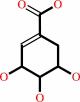 shikimate
|
+
|
 ATP
ATP
|
=
|
3-phosphoshikimate
|
+
|
ADP
Bound ligand (Het Group name = )
corresponds exactly
|
+
|
H(+)
|
|
|
|
|
|
|
|
|
|
|
|
|
Molecule diagrams generated from .mol files obtained from the
KEGG ftp site
|
|
|
|
|
|
|
|
|
|
|
|
|
|
 |
|
|
|
|
|
|
| |
|
|
| |
|
|
Acta Crystallogr Sect F Struct Biol Cryst Commun
63:1-6
(2007)
|
|
PubMed id:
|
|
|
|
|
|
| |
|
Effects of the magnesium and chloride ions and shikimate on the structure of shikimate kinase from Mycobacterium tuberculosis.
|
|
M.V.Dias,
L.M.Faím,
I.B.Vasconcelos,
J.S.de Oliveira,
L.A.Basso,
D.S.Santos,
W.F.de Azevedo.
|
|
|
|
|
| |
ABSTRACT
|
|
|
|
| |
|
|
Bacteria, fungi and plants can convert carbohydrate and phosphoenolpyruvate into
chorismate, which is the precursor of various aromatic compounds. The seven
enzymes of the shikimate pathway are responsible for this conversion. Shikimate
kinase (SK) is the fifth enzyme in this pathway and converts shikimate to
shikimate-3-phosphate. In this work, the conformational changes that occur on
binding of shikimate, magnesium and chloride ions to SK from Mycobacterium
tuberculosis (MtSK) are described. It was observed that both ions and shikimate
influence the conformation of residues of the active site of MtSK. Magnesium
influences the conformation of the shikimate hydroxyl groups and the position of
the side chains of some of the residues of the active site. Chloride seems to
influence the affinity of ADP and its position in the active site and the
opening length of the LID domain. Shikimate binding causes a closing of the LID
domain and also seems to influence the crystallographic packing of SK. The
results shown here could be useful for understanding the catalytic mechanism of
SK and the role of ions in the activity of this protein.
|
|
|
|
|
|
|
|
|
|
|
|
|
|
|
|
|
|
 |
 |
 |
|
|


 Links
Links


