 |
PDBsum entry 1seh

|
|
|
|
|
 |
Contents |
 |
|
|
|
|
|
|
|
|
|
|
|
|
* Residue conservation analysis
|
|
|
|
|
|
|
|
|
|
|
Enzyme class:
|
|
E.C.3.6.1.23
- dUTP diphosphatase.
|
|
|
|
|
|
|
Reaction:
|
|
dUTP + H2O = dUMP + diphosphate + H+
|
|
|
|
|
|
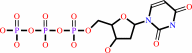 dUTP
dUTP
|
+
|
H2O
|
=
|
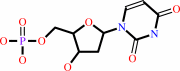 dUMP
dUMP
|
+
|
diphosphate
Bound ligand (Het Group name = )
corresponds exactly
|
+
|
H(+)
|
|
|
|
|
|
|
|
|
|
|
|
|
Molecule diagrams generated from .mol files obtained from the
KEGG ftp site
|
|
|
|
|
|
|
|
|
|
|
|
|
|
 |
|
|
|
|
|
|
| |
|
|
| |
|
DOI no:
|
J Biol Chem
279:42907-42915
(2004)
|
|
PubMed id:
|
|
|
|
|
|
| |
|
Structural insights into the catalytic mechanism of phosphate ester hydrolysis by dUTPase.
|
|
O.Barabás,
V.Pongrácz,
J.Kovári,
M.Wilmanns,
B.G.Vértessy.
|
|
|
|
|
| |
ABSTRACT
|
|
|
|
| |
|
|
dUTPase is essential to keep uracil out of DNA. Crystal structures of substrate
(dUTP and alpha,beta-imino-dUTP) and product complexes of wild type and mutant
dUTPases were determined to reveal how an enzyme responsible for DNA integrity
functions. A kinetic analysis of wild type and mutant dUTPases was performed to
obtain relevant mechanistic information in solution. Substrate hydrolysis is
shown to be initiated via in-line nucleophile attack of a water molecule
oriented by an activating conserved aspartate residue. Substrate binding in a
catalytically competent conformation is achieved by (i) multiple interactions of
the triphosphate moiety with catalysis-assisting Mg2+, (ii) a concerted motion
of residues from three conserved enzyme motifs as compared with the apoenzyme,
and (iii) an intricate hydrogen-bonding network that includes several water
molecules in the active site. Results provide an understanding for the catalytic
role of conserved residues in dUTPases.
|
|
|
|
|
|
| |
Selected figure(s)
|
|
|
|
| |
|
|

|
|

|
|
Figure 3.
FIG. 3. Identification of the nucleophile water. A,
simulated annealed omit electron density map, restricted to
exclusively show the exact position of the catalytic water
molecule in the wild type dUTPase:  , ,  -imino-dUTP:Mg2+
structure. The figure also shows the hydrogen-bonding network
involving the phosphate chain in this complex structure. In
addition to the catalytic water, Mg2+-coordinating waters, W1,
W2, W4, W15, and W21, also participate in the primary
hydrogen-bonding interactions. B, superimposed structures of
wild type (dark tones) and Asp90 -imino-dUTP:Mg2+
structure. The figure also shows the hydrogen-bonding network
involving the phosphate chain in this complex structure. In
addition to the catalytic water, Mg2+-coordinating waters, W1,
W2, W4, W15, and W21, also participate in the primary
hydrogen-bonding interactions. B, superimposed structures of
wild type (dark tones) and Asp90  Asn mutant (light tones)
dUTPase: , -imino-dUTP:Mg2+
complexes. Note that the only remarkable difference between the
superimposed structures is the disappearance of W[cat] from the
mutant complex. Atomic color code: carbon, dark/light gray;
oxygen, dark/light red (pink); phosphorus, dark/light orange
(yellow); nitrogen, dark/light blue; magnesium, dark/light
purple. C, superimposed structures of Asp90 Asn mutant dUTPase:
dUTP:Mg2+ (dark tones) and Asp90 Asn mutant dUTPase: , -imino-dUTP:Mg2+ (light
tones) complexes. Note the close identity in the positions of
the nucleotide ligands. D, apoenzyme retains a water closely
corresponding to the W[cat] position. 3-Fold superimposition of
the apoenzyme (green carbons and water, otherwise standard atom
coloring), enzyme-substrate (dark tones), and enzyme-product
(light tones) structures. Note the position of the catalytic
water from the apoenzyme to the enzyme-substrate and
enzyme-product complexes. E, F, and G, simulated annealed omit
electron density maps for the substrates in wild type E. coli
dUTPase: , -imino-dUTP:Mg2+, the
Asp90 Asn E. coli dUTPase:
,
-imino-dUTP: Mg2+, and
the Asp90 Asn E. coli
dUTPase:dUTP:Mg2+ structures, respectively. Maps are restricted
to show the nucleotide ligand, the Mg2+, the three water
molecules coordinating to the metal ion, as well as the
catalytic water, if present. Asn mutant (light tones)
dUTPase: , -imino-dUTP:Mg2+
complexes. Note that the only remarkable difference between the
superimposed structures is the disappearance of W[cat] from the
mutant complex. Atomic color code: carbon, dark/light gray;
oxygen, dark/light red (pink); phosphorus, dark/light orange
(yellow); nitrogen, dark/light blue; magnesium, dark/light
purple. C, superimposed structures of Asp90 Asn mutant dUTPase:
dUTP:Mg2+ (dark tones) and Asp90 Asn mutant dUTPase: , -imino-dUTP:Mg2+ (light
tones) complexes. Note the close identity in the positions of
the nucleotide ligands. D, apoenzyme retains a water closely
corresponding to the W[cat] position. 3-Fold superimposition of
the apoenzyme (green carbons and water, otherwise standard atom
coloring), enzyme-substrate (dark tones), and enzyme-product
(light tones) structures. Note the position of the catalytic
water from the apoenzyme to the enzyme-substrate and
enzyme-product complexes. E, F, and G, simulated annealed omit
electron density maps for the substrates in wild type E. coli
dUTPase: , -imino-dUTP:Mg2+, the
Asp90 Asn E. coli dUTPase:
,
-imino-dUTP: Mg2+, and
the Asp90 Asn E. coli
dUTPase:dUTP:Mg2+ structures, respectively. Maps are restricted
to show the nucleotide ligand, the Mg2+, the three water
molecules coordinating to the metal ion, as well as the
catalytic water, if present.
|
|
Figure 4.
FIG. 4. Interaction mapping in enzyme-substrate (A), and
enzyme-product complexes (B). Interactions are shown only for
the phosphate chain moiety of the ligand. Due to the close
similarity of the nucleotide interactions in the three
enzyme-substrate complexes determined in the present study (cf.
Fig. 3 and Table I), the map was selected to show the actual
distances as found in the wild type dUTPase: , -imino-dUTP: Mg2+ (X =
N) complex where W[cat] is also present. In the Asp90 Asn
mutant dUTPase: , -imino-dUTP:Mg2+ (X = N)
and Asp90 Asn mutant dUTPase:dUTP:
Mg2+ (X = O) complex, the only significant differences are that
(i) W[cat] is absent and Asp90O  2 becomes AsnN 2 and
(ii) in the Asp90 Asn mutant dUTPase:dUTP:
Mg2+ (X = O) complex, the X-Ser72O 2 becomes AsnN 2 and
(ii) in the Asp90 Asn mutant dUTPase:dUTP:
Mg2+ (X = O) complex, the X-Ser72O  interaction is absent.
Changes in all other distances are within ±0.2 Å. interaction is absent.
Changes in all other distances are within ±0.2 Å.
|
|
|
|
|
|
| |
The above figures are
reprinted
by permission from the ASBMB:
J Biol Chem
(2004,
279,
42907-42915)
copyright 2004.
|
|
| |
Figures were
selected
by an automated process.
|
|
|
|
|
|
|
|
|
|
|
|
|
|
|
|
 |
 |
 |
|
Literature references that cite this PDB file's key reference
|
|
|
| |
PubMed id
|
|
Reference
|
|
|
|
|
|
I.Pecsi,
I.Leveles,
V.Harmat,
B.G.Vertessy,
and
J.Toth
(2010).
Aromatic stacking between nucleobase and enzyme promotes phosphate ester hydrolysis in dUTPase.
|
| |
Nucleic Acids Res,
38,
7179-7186.
|
|
|
PDB codes:
|
|
|
|
|
|
|
|
J.García-Nafría,
L.Burchell,
M.Takezawa,
N.J.Rzechorzek,
M.J.Fogg,
and
K.S.Wilson
(2010).
The structure of the genomic Bacillus subtilis dUTPase: novel features in the Phe-lid.
|
| |
Acta Crystallogr D Biol Crystallogr,
66,
953-961.
|
|
|
PDB codes:
|
|
|
|
|
|
|
|
B.G.Vértessy,
and
J.Tóth
(2009).
Keeping uracil out of DNA: physiological role, structure and catalytic mechanism of dUTPases.
|
| |
Acc Chem Res,
42,
97.
|
|
|
|
|
|
|
G.L.Li,
J.Wang,
L.F.Li,
and
X.D.Su
(2009).
Crystallization and preliminary X-ray analysis of three dUTPases from Gram-positive bacteria.
|
| |
Acta Crystallogr Sect F Struct Biol Cryst Commun,
65,
339-342.
|
|
|
|
|
|
|
L.Freeman,
M.Buisson,
N.Tarbouriech,
A.Van der Heyden,
P.Labbé,
and
W.P.Burmeister
(2009).
The flexible motif V of Epstein-Barr virus deoxyuridine 5'-triphosphate pyrophosphatase is essential for catalysis.
|
| |
J Biol Chem,
284,
25280-25289.
|
|
|
PDB codes:
|
|
|
|
|
|
|
|
J.Kovári,
O.Barabás,
B.Varga,
A.Békési,
F.Tölgyesi,
J.Fidy,
J.Nagy,
and
B.G.Vértessy
(2008).
Methylene substitution at the alpha-beta bridging position within the phosphate chain of dUDP profoundly perturbs ligand accommodation into the dUTPase active site.
|
| |
Proteins,
71,
308-319.
|
|
|
PDB codes:
|
|
|
|
|
|
|
|
A.Samal,
N.Schormann,
W.J.Cook,
L.J.DeLucas,
and
D.Chattopadhyay
(2007).
Structures of vaccinia virus dUTPase and its nucleotide complexes.
|
| |
Acta Crystallogr D Biol Crystallogr,
63,
571-580.
|
|
|
PDB codes:
|
|
|
|
|
|
|
|
I.Berente,
E.Czinki,
and
G.Náray-Szabó
(2007).
A combined electronegativity equalization and electrostatic potential fit method for the determination of atomic point charges.
|
| |
J Comput Chem,
28,
1936-1942.
|
|
|
|
|
|
|
V.Németh-Pongrácz,
O.Barabás,
M.Fuxreiter,
I.Simon,
I.Pichová,
M.Rumlová,
H.Zábranská,
D.Svergun,
M.Petoukhov,
V.Harmat,
E.Klement,
E.Hunyadi-Gulyás,
K.F.Medzihradszky,
E.Kónya,
and
B.G.Vértessy
(2007).
Flexible segments modulate co-folding of dUTPase and nucleocapsid proteins.
|
| |
Nucleic Acids Res,
35,
495-505.
|
|
|
PDB codes:
|
|
|
|
|
|
|
|
A.Guranowski,
E.Starzyńska,
M.Pietrowska-Borek,
J.Jemielity,
J.Kowalska,
E.Darzynkiewicz,
M.J.Thompson,
and
G.M.Blackburn
(2006).
Methylene analogues of adenosine 5'-tetraphosphate. Their chemical synthesis and recognition by human and plant mononucleoside tetraphosphatases and dinucleoside tetraphosphatases.
|
| |
FEBS J,
273,
829-838.
|
|
|
|
|
|
|
M.Guillet,
P.A.Van Der Kemp,
and
S.Boiteux
(2006).
dUTPase activity is critical to maintain genetic stability in Saccharomyces cerevisiae.
|
| |
Nucleic Acids Res,
34,
2056-2066.
|
|
|
|
|
|
|
S.U.Lari,
C.Y.Chen,
B.G.Vertéssy,
J.Morré,
and
S.E.Bennett
(2006).
Quantitative determination of uracil residues in Escherichia coli DNA: Contribution of ung, dug, and dut genes to uracil avoidance.
|
| |
DNA Repair (Amst),
5,
1407-1420.
|
|
|
|
|
|
|
N.Tarbouriech,
M.Buisson,
J.M.Seigneurin,
S.Cusack,
and
W.P.Burmeister
(2005).
The monomeric dUTPase from Epstein-Barr virus mimics trimeric dUTPases.
|
| |
Structure,
13,
1299-1310.
|
|
|
PDB codes:
|
|
|
|
|
|
|
The most recent references are shown first.
Citation data come partly from CiteXplore and partly
from an automated harvesting procedure. Note that this is likely to be
only a partial list as not all journals are covered by
either method. However, we are continually building up the citation data
so more and more references will be included with time.
Where a reference describes a PDB structure, the PDB
codes are
shown on the right.
|
|


 Links
Links


