 |
PDBsum entry 1nug

|
|
|
|
|
 |
Contents |
 |
|
|
|
|
|
|
|
|
|
|
|
|
* Residue conservation analysis
|
|
|
|
|
|
|
|
|
|
|
Enzyme class:
|
|
E.C.2.3.2.13
- protein-glutamine gamma-glutamyltransferase.
|
|
|
|
|
|
|
Reaction:
|
|
L-glutaminyl-[protein] + L-lysyl-[protein] = [protein]-L-lysyl-N6-5- L-glutamyl-[protein] + NH4+
|
|
|
|
|
|
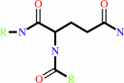 protein-L-glutamine
|
+
|
protein-L-lysine
|
=
|
protein with an N(6)- (gamma-glutamyl)-L-lysine cross-link
|
+
|
NH(3)
|
|
|
|
|
|
|
|
|
|
Cofactor:
|
|
Ca(2+)
|
|
|
|
|
|
|
|
|
Molecule diagrams generated from .mol files obtained from the
KEGG ftp site
|
|
|
|
|
|
|
|
|
|
|
|
|
|
 |
|
|
|
|
|
|
| |
|
|
| |
|
DOI no:
|
J Biol Chem
278:23834-23841
(2003)
|
|
PubMed id:
|
|
|
|
|
|
| |
|
Roles of calcium ions in the activation and activity of the transglutaminase 3 enzyme.
|
|
B.Ahvazi,
K.M.Boeshans,
W.Idler,
U.Baxa,
P.M.Steinert.
|
|
|
|
|
| |
ABSTRACT
|
|
|
|
| |
|
|
The transglutaminase 3 enzyme is widely expressed in many tissues including
epithelia. We have shown previously that it can bind three Ca2+ ions, which in
site one is constitutively bound, while those in sites two and three are
acquired during activation and are required for activity. In particular, binding
at site three opens a channel through the enzyme and exposes two tryptophan
residues near the active site that are thought to be important for enzyme
reaction. In this study, we have solved the structures of three more forms of
this enzyme by x-ray crystallography in the presence of Ca2+ and/or Mg2+, which
provide new insights on the precise contribution of each Ca2+ ion to activation
and activity. First, we found that Ca2+ ion in site one can be exchanged with
difficulty, and it has a binding affinity of Kd = 0.3 microm (DeltaH = -6.70 +/-
0.52 kcal/mol), which suggests it is important for the stabilization of the
enzyme. Site two can be occupied by some lanthanides but only Ca2+ of the Group
2 family of alkali earth metals, and its occupancy are required for activity.
Site three can be occupied by some lanthanides, Ca2+,or Mg2+; however, when Mg2+
is present, the enzyme is inactive, and the channel is closed. Thus Ca2+ binding
in both sites two and three cooperate in opening the channel. We speculate that
manipulation of the channel opening could be controlled by intracellular cation
levels. Together, these data have important implications for reaction mechanism
of the enzyme: the opening of a channel perhaps controls access to and
manipulation of substrates at the active site.
|
|
|
|
|
|
| |
Selected figure(s)
|
|
|
|
| |
|
|

|
|

|
|
Figure 1.
FIG. 1. Conformations of the forms I (a and b), II (c and
d), and III (e and g) solved in this study. The upper row shows
the solved structures of the three forms. This is nominally the
front side of the enzyme. The amino-terminal  -sandwich (red),
catalytic core (blue), -barrel 1 (magenta), and
-barrel 2 (orange)
domains are shown. The Ca^2^+ ions are shown in yellow, the sole
Mg2^+ ion in cyan. Below are shown the electrostatic surface
potential images. The acidic and basic residues are colored red
and blue, respectively. The electrostatic potentials, including
Ca^2^+ and Mg2^+ ions, have been mapped onto the surface plan
from -15 kT (deep red) to +15 kT (deep blue). The open channel
is clearly evident in b. In g, the back side of the enzyme has a
deep cavity; the front side (f) remains closed. -sandwich (red),
catalytic core (blue), -barrel 1 (magenta), and
-barrel 2 (orange)
domains are shown. The Ca^2^+ ions are shown in yellow, the sole
Mg2^+ ion in cyan. Below are shown the electrostatic surface
potential images. The acidic and basic residues are colored red
and blue, respectively. The electrostatic potentials, including
Ca^2^+ and Mg2^+ ions, have been mapped onto the surface plan
from -15 kT (deep red) to +15 kT (deep blue). The open channel
is clearly evident in b. In g, the back side of the enzyme has a
deep cavity; the front side (f) remains closed.
|
|
Figure 2.
FIG. 2. Identification of key residues involved in the
coordination with metal ions in sites one, two, and three in
forms I-III. The details of these interactions and the bond
lengths are summarized in Table III.
|
|
|
|
|
|
| |
The above figures are
reprinted
by permission from the ASBMB:
J Biol Chem
(2003,
278,
23834-23841)
copyright 2003.
|
|
| |
Figures were
selected
by an automated process.
|
|
|
|
|
|
|
|
|
|
|
|
|
|
|
|
 |
 |
 |
|
Literature references that cite this PDB file's key reference
|
|
|
| |
PubMed id
|
|
Reference
|
|
|
|
|
|
A.Yamane,
M.Fukui,
Y.Sugimura,
M.Itoh,
M.P.Alea,
V.Thomas,
S.El Alaoui,
M.Akiyama,
and
K.Hitomi
(2010).
Identification of a preferred substrate peptide for transglutaminase 3 and detection of in situ activity in skin and hair follicles.
|
| |
FEBS J,
277,
3564-3574.
|
|
|
|
|
|
|
H.H.Bragulla,
and
D.G.Homberger
(2009).
Structure and functions of keratin proteins in simple, stratified, keratinized and cornified epithelia.
|
| |
J Anat,
214,
516-559.
|
|
|
|
|
|
|
K.Aufenvenne,
V.Oji,
T.Walker,
C.Becker-Pauly,
H.C.Hennies,
W.Stöcker,
and
H.Traupe
(2009).
Transglutaminase-1 and bathing suit ichthyosis: molecular analysis of gene/environment interactions.
|
| |
J Invest Dermatol,
129,
2068-2071.
|
|
|
|
|
|
|
T.M.Jeitner,
N.A.Muma,
K.P.Battaile,
and
A.J.Cooper
(2009).
Transglutaminase activation in neurodegenerative diseases.
|
| |
Future Neurol,
4,
449-467.
|
|
|
|
|
|
|
V.Pietroni,
S.Di Giorgi,
A.Paradisi,
B.Ahvazi,
E.Candi,
and
G.Melino
(2008).
Inactive and highly active, proteolytically processed transglutaminase-5 in epithelial cells.
|
| |
J Invest Dermatol,
128,
2760-2766.
|
|
|
|
|
|
|
K.M.Boeshans,
T.C.Mueser,
and
B.Ahvazi
(2007).
A three-dimensional model of the human transglutaminase 1: insights into the understanding of lamellar ichthyosis.
|
| |
J Mol Model,
13,
233-246.
|
|
|
|
|
|
|
M.T.Sturniolo,
R.A.Chandraratna,
and
R.L.Eckert
(2005).
A novel transglutaminase activator forms a complex with type 1 transglutaminase.
|
| |
Oncogene,
24,
2963-2972.
|
|
|
|
|
|
|
R.L.Eckert,
M.T.Sturniolo,
A.M.Broome,
M.Ruse,
and
E.A.Rorke
(2005).
Transglutaminase function in epidermis.
|
| |
J Invest Dermatol,
124,
481-492.
|
|
|
|
|
|
The most recent references are shown first.
Citation data come partly from CiteXplore and partly
from an automated harvesting procedure. Note that this is likely to be
only a partial list as not all journals are covered by
either method. However, we are continually building up the citation data
so more and more references will be included with time.
|
|


 Links
Links


