5fp6

X-ray diffraction
1.85Å resolution

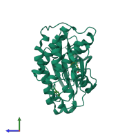
Structure of cyclin-dependent kinase 2 with small-molecule ligand 3-(4,7-dichloro-1H-indol-3-yl)prop-2-yn-1-ol (AT17833) in an alternate binding site.

Released:
Source organism: Homo sapiens
Primary publication:
Detection of secondary binding sites in proteins using fragment screening.
Proc Natl Acad Sci U S A 112 15910-5 (2015)
PMID: 26655740

Function and Biology Details

Structure analysis Details

Assembly composition:
monomeric (preferred)
PDBe Complex ID:
PDB-CPX-150313 (preferred)
Entry contents:
1 distinct polypeptide molecule
Macromolecule:
Cyclin-dependent kinase 2 Chain: A
Molecule details ›
Chain: A
Length: 298 amino acids
Theoretical weight: 33.98 KDa
Source organism: Homo sapiens
Expression system: Spodoptera frugiperda
UniProt:
  • Canonical: P24941 (Residues: 1-298; Coverage: 100%)
Gene names: CDK2, CDKN2
Sequence domains: Protein kinase domain
Structure domains:

Ligands and Environments

1 bound ligand:
No modified residues

Experiments and Validation Details

Entry percentile scores
X-ray source: RIGAKU FR-E
Spacegroup: P212121
Unit cell:
a: 53.917Å b: 72.214Å c: 72.198Å
α: 90° β: 90° γ: 90°
R-values:
R R work R free
0.209 0.207 0.24
Expression system: Spodoptera frugiperda