5cew

X-ray diffraction
2.03Å resolution

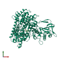
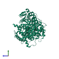
Crystal structure of Mycobacterium tuberculosis malate synthase in complex with 2-(pyridin-4-yl)thiazolidine-4-carboxylic acid

Released:

Function and Biology Details

Reaction catalysed:
Acetyl-CoA + H(2)O + glyoxylate = (S)-malate + CoA
Biochemical function:
Biological process:
Cellular component:

Structure analysis Details

Assembly composition:
monomeric (preferred)
Assembly name:
PDBe Complex ID:
PDB-CPX-161718 (preferred)
Entry contents:
1 distinct polypeptide molecule
Macromolecule:
Malate synthase G Chain: A
Molecule details ›
Chain: A
Length: 741 amino acids
Theoretical weight: 80.46 KDa
Source organism: Mycobacterium tuberculosis H37Rv
Expression system: Escherichia coli BL21
UniProt:
  • Canonical: P9WK17 (Residues: 1-741; Coverage: 100%)
Gene names: MTCY1A11.06, Rv1837c, glcB
Sequence domains:
Structure domains:

Ligands and Environments

2 bound ligands:
No modified residues

Experiments and Validation Details

Entry percentile scores
X-ray source: APS BEAMLINE 23-ID-D
Spacegroup: P43212
Unit cell:
a: 78.379Å b: 78.379Å c: 225.187Å
α: 90° β: 90° γ: 90°
R-values:
R R work R free
0.209 0.205 0.292
Expression system: Escherichia coli BL21