4pg5

X-ray diffraction
2.2Å resolution

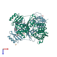
Crystal structure of S. aureus Homoserine Dehydrogenase at pH6.5

Released:
Source organism: Staphylococcus aureus M1064
Primary publication:
Structural basis for the catalytic mechanism of homoserine dehydrogenase.
Acta Crystallogr D Biol Crystallogr 71 1216-25 (2015)
PMID: 25945586

Function and Biology Details

Reaction catalysed:
L-homoserine + NAD(P)(+) = L-aspartate 4-semialdehyde + NAD(P)H
Biochemical function:
Biological process:
Cellular component:
  • not assigned

Structure analysis Details

Assembly composition:
homo dimer (preferred)
PDBe Complex ID:
PDB-CPX-102390 (preferred)
Entry contents:
1 distinct polypeptide molecule
Macromolecule:
Homoserine dehydrogenase Chains: A, B
Molecule details ›
Chains: A, B
Length: 468 amino acids
Theoretical weight: 51.38 KDa
Source organism: Staphylococcus aureus M1064
Expression system: Escherichia coli
UniProt:
  • Canonical: A0A0M3KKV6 (Residues: 1-426; Coverage: 95%)
Gene name: U5K_01898
Sequence domains:
Structure domains:

Ligands and Environments

No modified residues

Experiments and Validation Details

Entry percentile scores
X-ray source: ESRF BEAMLINE BM14
Spacegroup: P212121
Unit cell:
a: 72.48Å b: 115.43Å c: 118.23Å
α: 90° β: 90° γ: 90°
R-values:
R R work R free
0.203 0.201 0.238
Expression system: Escherichia coli