4o79

X-ray diffraction
2Å resolution

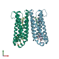
Crystal Structure of Ascorbate-bound Cytochrome b561, crystal soaked in 1 M L-ascorbate for 10 minutes

Released:
Source organism: Arabidopsis thaliana
Primary publication:
Structure and mechanism of a eukaryotic transmembrane ascorbate-dependent oxidoreductase.
Proc Natl Acad Sci U S A 111 1813-8 (2014)
PMID: 24449903

Function and Biology Details

Reaction catalysed:
Ascorbate(Side 1) + Fe(III)(Side 2) = monodehydroascorbate(Side 1) + Fe(II)(Side 2)
Biological process:
Cellular component:

Structure analysis Details

Assembly composition:
homo dimer (preferred)
PDBe Complex ID:
PDB-CPX-193555 (preferred)
Entry contents:
1 distinct polypeptide molecule
Macromolecule:
Transmembrane ascorbate ferrireductase 2 Chains: A, B
Molecule details ›
Chains: A, B
Length: 230 amino acids
Theoretical weight: 25.31 KDa
Source organism: Arabidopsis thaliana
Expression system: Escherichia coli
UniProt:
  • Canonical: Q9SWS1 (Residues: 1-230; Coverage: 100%)
Gene names: ACYB-1, At5g38630, CYB-1, CYB561B, CYB561B2, CYBASC2, CYTB561, MBB18.18
Sequence domains: Eukaryotic cytochrome b561
Structure domains: Four Helix Bundle (Hemerythrin (Met), subunit A)

Ligands and Environments

3 bound ligands:
No modified residues

Experiments and Validation Details

Entry percentile scores
X-ray source: SSRF BEAMLINE BL17U
Spacegroup: I222
Unit cell:
a: 73.193Å b: 108.648Å c: 111.389Å
α: 90° β: 90° γ: 90°
R-values:
R R work R free
0.202 0.202 0.213
Expression system: Escherichia coli