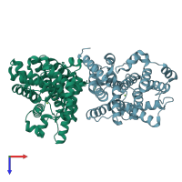
4ajf

X-ray diffraction
1.9Å resolution

Identification and structural characterization of PDE10 fragment inhibitors

Released:
Source organism: Homo sapiens
Entry authors: Johansson P, Albert JS, Spadola L, Akerud T, Back E, Hillertz P, Horsefeld R, Scott C, Spear N, Tian G, Tigerstrom A, Aharony D, Geschwindner S

Function and Biology Details

Structure analysis Details

Assembly composition:
homo dimer (preferred)
PDBe Complex ID:
PDB-CPX-195089 (preferred)
Entry contents:
1 distinct polypeptide molecule
Macromolecule:
cAMP and cAMP-inhibited cGMP 3',5'-cyclic phosphodiesterase 10A Chains: A, D
Molecule details ›
Chains: A, D
Length: 328 amino acids
Theoretical weight: 37.93 KDa
Source organism: Homo sapiens
Expression system: Escherichia coli
UniProt:
  • Canonical: Q9Y233 (Residues: 715-1040; Coverage: 31%)
Gene name: PDE10A
Structure domains: 3'5'-cyclic nucleotide phosphodiesterase, catalytic domain

Ligands and Environments

3 bound ligands:
No modified residues

Experiments and Validation Details

Entry percentile scores
X-ray source: RIGAKU FR-E
Spacegroup: P212121
Unit cell:
a: 49.371Å b: 81.798Å c: 156.596Å
α: 90° β: 90° γ: 90°
R-values:
R R work R free
0.215 0.213 0.248
Expression system: Escherichia coli