3kb2

X-ray diffraction
2.2Å resolution

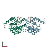
Crystal Structure of YorR protein in complex with phosphorylated GDP from Bacillus subtilis, Northeast Structural Genomics Consortium Target SR256

Released:
Source organism: Bacillus subtilis
Entry authors: Forouhar F, Friedman D, Seetharaman J, Janjua J, Xiao R, Cunningham K, Ma L, Ho C, Everett JK, Nair R, Acton TB, Rost B, Montelione GT, Tong L, Hunt JF, Northeast Structural Genomics Consortium (NESG)

Function and Biology Details

Biochemical function:
Cellular component:

Structure analysis Details

Assembly composition:
homo dimer (preferred)
PDBe Complex ID:
PDB-CPX-128389 (preferred)
Entry contents:
1 distinct polypeptide molecule
Macromolecule:
SPBc2 prophage-derived uncharacterized protein yorR Chains: A, B
Molecule details ›
Chains: A, B
Length: 173 amino acids
Theoretical weight: 20.26 KDa
Source organism: Bacillus subtilis
Expression system: Escherichia coli BL21(DE3)
UniProt:
  • Canonical: O31896 (Residues: 1-165; Coverage: 100%)
Gene names: BSU20280, yorR
Structure domains: P-loop containing nucleotide triphosphate hydrolases

Ligands and Environments

2 bound ligands:
1 modified residue:

Experiments and Validation Details

Entry percentile scores
X-ray source: NSLS BEAMLINE X4A
Spacegroup: P21
Unit cell:
a: 45.478Å b: 63.05Å c: 70.456Å
α: 90° β: 91.57° γ: 90°
R-values:
R R work R free
0.197 0.195 0.262
Expression system: Escherichia coli BL21(DE3)