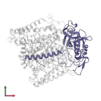
Assemblies
Assembly Name:
Reaction Centre
Multimeric state:
hetero trimer
Accessible surface area:
29236.15 Å2
Buried surface area:
37111.94 Å2
Dissociation area:
926.12
Å2
Dissociation energy (ΔGdiss):
9.47
kcal/mol
Dissociation entropy (TΔSdiss):
7.76
kcal/mol
Symmetry number:
1
PDBe Complex ID:
PDB-CPX-142934
Macromolecules
Chain: L
Length: 281 amino acids
Theoretical weight: 31.35 KDa
Source organism: Cereibacter sphaeroides
Expression system: Cereibacter sphaeroides
UniProt:
Pfam: Photosynthetic reaction centre protein
InterPro:
Length: 281 amino acids
Theoretical weight: 31.35 KDa
Source organism: Cereibacter sphaeroides
Expression system: Cereibacter sphaeroides
UniProt:
- Canonical:
 P0C0Y8 (Residues: 2-282; Coverage: 100%)
Pfam: Photosynthetic reaction centre protein
InterPro:
- Photosystem II protein D1/D2 superfamily
- Photosynthetic reaction centre, L/M
- Photosynthetic reaction centre, L subunit
Chain: M
Length: 314 amino acids
Theoretical weight: 35.37 KDa
Source organism: Cereibacter sphaeroides
Expression system: Cereibacter sphaeroides
UniProt:
Pfam: Photosynthetic reaction centre protein
InterPro:
Length: 314 amino acids
Theoretical weight: 35.37 KDa
Source organism: Cereibacter sphaeroides
Expression system: Cereibacter sphaeroides
UniProt:
- Canonical:
 P0C0Y9 (Residues: 2-308; Coverage: 100%)
Pfam: Photosynthetic reaction centre protein
InterPro:
- Photosystem II protein D1/D2 superfamily
- Photosynthetic reaction centre, L/M
- Photosynthetic reaction centre, M subunit
Chain: H
Length: 260 amino acids
Theoretical weight: 28.07 KDa
Source organism: Cereibacter sphaeroides
Expression system: Cereibacter sphaeroides
UniProt:
Pfam:
InterPro:
Length: 260 amino acids
Theoretical weight: 28.07 KDa
Source organism: Cereibacter sphaeroides
Expression system: Cereibacter sphaeroides
UniProt:
- Canonical:
 P0C0Y7 (Residues: 1-260; Coverage: 100%)
Pfam:
InterPro:
- Photosynthetic reaction centre, H subunit
- Photosynthetic reaction centre, H subunit, N-terminal
- Photosynthetic reaction centre, H subunit, N-terminal domain superfamily
- PRC-barrel-like superfamily
- Bacterial photosynthetic reaction centre, H-chain, C-terminal
- PRC-barrel domain


