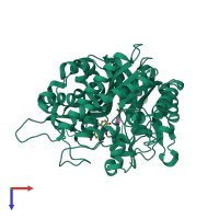
3aiw

X-ray diffraction
2.4Å resolution

Crystal structure of beta-glucosidase in rye complexed with 2-deoxy-2-fluoroglucoside and dinitrophenol

Released:
Source organism: Secale cereale

Function and Biology Details

Reactions catalysed:
(2R)-4-hydroxy-3-oxo-3,4-dihydro-2H-1,4-benzoxazin-2-yl beta-D-glucopyranoside + H(2)O = 2,4-dihydroxy-2H-1,4-benzoxazin-3(4H)-one + D-glucose
Hydrolysis of terminal, non-reducing beta-D-glucosyl residues with release of beta-D-glucose
Biological process:
Cellular component:

Structure analysis Details

Assemblies composition:
homo hexamer
homo dimer (preferred)
PDBe Complex ID:
PDB-CPX-190385 (preferred)
Entry contents:
1 distinct polypeptide molecule
Macromolecule:
4-hydroxy-7-methoxy-3-oxo-3,4-dihydro-2H-1,4-benzoxazin-2-yl glucoside beta-D-glucosidase, chloroplastic Chain: A
Molecule details ›
Chain: A
Length: 564 amino acids
Theoretical weight: 63.97 KDa
Source organism: Secale cereale
Expression system: Escherichia coli
UniProt:
  • Canonical: Q9FYS3 (Residues: 50-568; Coverage: 91%)
Sequence domains: Glycosyl hydrolase family 1
Structure domains: Glycosidases

Ligands and Environments

2 bound ligands:
No modified residues

Experiments and Validation Details

Entry percentile scores
X-ray source: PHOTON FACTORY BEAMLINE BL-6A
Spacegroup: P4132
Unit cell:
a: 194.686Å b: 194.686Å c: 194.686Å
α: 90° β: 90° γ: 90°
R-values:
R R work R free
0.187 0.186 0.212
Expression system: Escherichia coli