


Function and Biology Details
Biochemical function:
Biological process:
Cellular component:
Sequence domains:
- Small GTPase
- P-loop containing nucleoside triphosphate hydrolase
- GMP phosphodiesterase, delta subunit
- Immunoglobulin E-set
- Retinal rod rhodopsin-sensitive cGMP 3', 5'-cyclic phosphodiesterase, delta subunit
- GMP phosphodiesterase, delta subunit superfamily
- Small GTPase, Ras-type
- Small GTP-binding protein domain
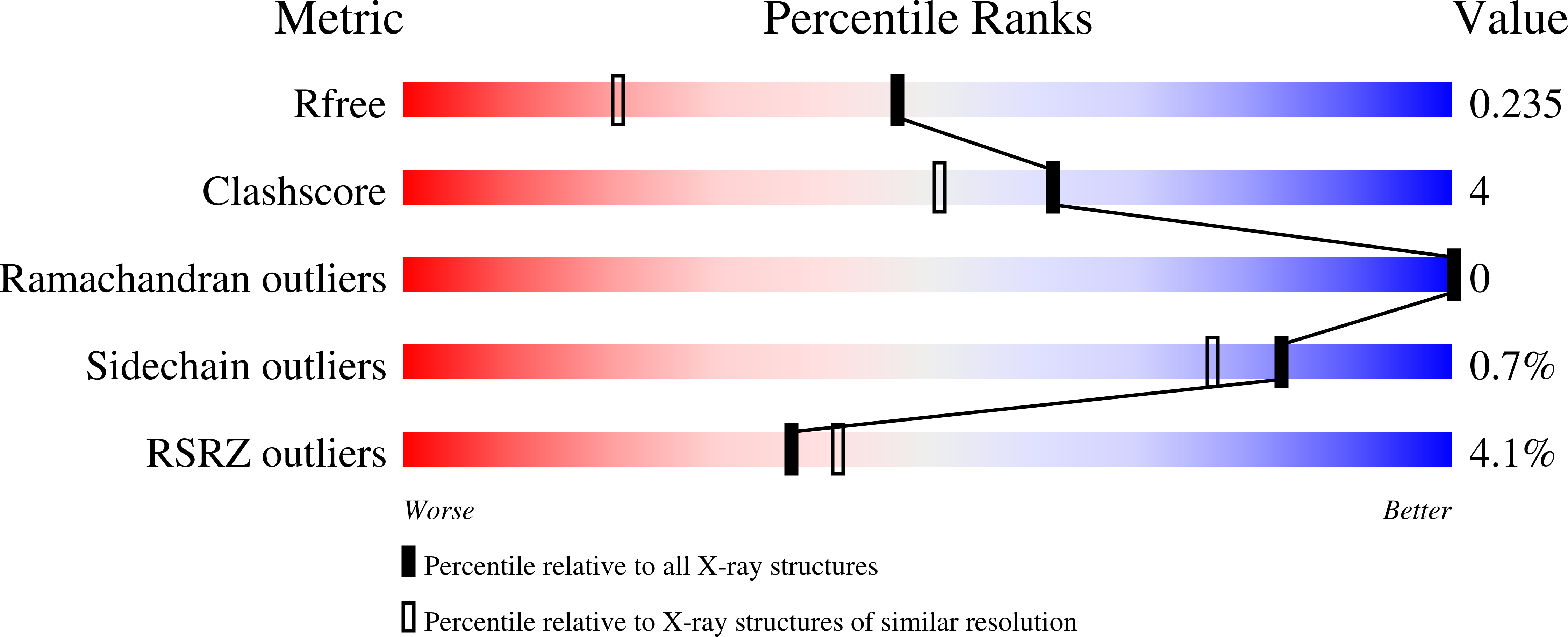
Structure analysis Details
Assembly composition:
hetero dimer (preferred)
Assembly name:
PDBe Complex ID:
PDB-CPX-129178 (preferred)
Entry contents:
2 distinct polypeptide molecules
Macromolecules (2 distinct):
GTP-binding protein Rheb
Molecule details ›
Chain: A
Length: 181 amino acids
Theoretical weight: 20.25 KDa
Source organism: Homo sapiens
Expression system: Escherichia coli
UniProt:
Sequence domains: Ras family
Structure domains: P-loop containing nucleotide triphosphate hydrolases
Length: 181 amino acids
Theoretical weight: 20.25 KDa
Source organism: Homo sapiens
Expression system: Escherichia coli
UniProt:
- Canonical:
 Q15382 (Residues: 1-181; Coverage: 98%)
Sequence domains: Ras family
Structure domains: P-loop containing nucleotide triphosphate hydrolases
Retinal rod rhodopsin-sensitive cGMP 3',5'-cyclic phosphodiesterase subunit delta
Molecule details ›
Chain: B
Length: 152 amino acids
Theoretical weight: 17.59 KDa
Source organism: Homo sapiens
Expression system: Escherichia coli
UniProt:
Sequence domains: GMP-PDE, delta subunit
Structure domains: GMP phosphodiesterase, delta subunit
Length: 152 amino acids
Theoretical weight: 17.59 KDa
Source organism: Homo sapiens
Expression system: Escherichia coli
UniProt:
- Canonical: O43924 (Residues: 1-150; Coverage: 100%)
Sequence domains: GMP-PDE, delta subunit
Structure domains: GMP phosphodiesterase, delta subunit


