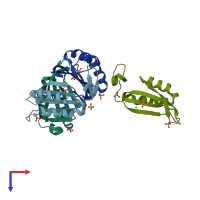
2z0g

X-ray diffraction
2.1Å resolution

The crystal structure of PII protein

Released:
Source organism: Aquifex aeolicus
Entry authors: Sakai H, Shinkai A, Kitamura Y, Kuramitsu S, Yokoyama S, RIKEN Structural Genomics/Proteomics Initiative (RSGI)

Function and Biology Details

Structure analysis Details

Assembly composition:
homo trimer (preferred)
PDBe Complex ID:
PDB-CPX-130325 (preferred)
Entry contents:
1 distinct polypeptide molecule
Macromolecule:
Nitrogen regulatory protein P-II Chains: A, B, C, D
Molecule details ›
Chains: A, B, C, D
Length: 112 amino acids
Theoretical weight: 12.52 KDa
Source organism: Aquifex aeolicus
Expression system: Escherichia coli
UniProt:
  • Canonical: O66513 (Residues: 1-112; Coverage: 100%)
Gene names: aq_109, glnB
Sequence domains: Nitrogen regulatory protein P-II
Structure domains: Alpha-Beta Plaits

Ligands and Environments

2 bound ligands:
No modified residues

Experiments and Validation Details

Entry percentile scores
X-ray source: SPRING-8 BEAMLINE BL26B2
Spacegroup: R3
Unit cell:
a: 114.398Å b: 114.398Å c: 70.067Å
α: 90° β: 90° γ: 120°
R-values:
R R work R free
0.186 0.186 0.23
Expression system: Escherichia coli