Function and Biology Details
Reactions catalysed:
Deoxynucleoside triphosphate + DNA(n) = diphosphate + DNA(n+1)
3'-end directed exonucleolytic cleavage of viral RNA-DNA hybrid
Endohydrolysis of RNA in RNA/DNA hybrids. Three different cleavage modes: 1. sequence-specific internal cleavage of RNA. Human immunodeficiency virus type 1 and Moloney murine leukemia virus enzymes prefer to cleave the RNA strand one nucleotide away from the RNA-DNA junction. 2. RNA 5'-end directed cleavage 13-19 nucleotides from the RNA end. 3. DNA 3'-end directed cleavage 15-20 nucleotides away from the primer terminus.
Endopeptidase for which the P1 residue is preferably hydrophobic.
Biochemical function:
Biological process:
Cellular component:
- not assigned
Structure analysis Details
Assembly composition:
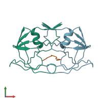
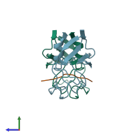
hetero trimer (preferred)
Assembly name:
Integrase and peptide (preferred)
PDBe Complex ID:
PDB-CPX-138000 (preferred)
Entry contents:
2 distinct polypeptide molecules
Macromolecules (2 distinct):
Ligands and Environments
No bound ligands
No modified residues
Experiments and Validation Details
Spacegroup:
P21
Expression systems:
- Escherichia coli
- Not provided


