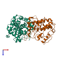
1jow

X-ray diffraction
3.1Å resolution

Crystal structure of a complex of human CDK6 and a viral cyclin

Released:
Primary publication:
Structural basis for CDK6 activation by a virus-encoded cyclin.
Nat Struct Biol 9 177-81 (2002)
PMID: 11828325

Function and Biology Details

Structure analysis Details

Assembly composition:
hetero dimer (preferred)
PDBe Complex ID:
PDB-CPX-169440 (preferred)
Entry contents:
2 distinct polypeptide molecules
Macromolecules (2 distinct):
Cyclin homolog Chain: A
Molecule details ›
Chain: A
Length: 254 amino acids
Theoretical weight: 28.67 KDa
Source organism: Saimiriine gammaherpesvirus 2
Expression system: Spodoptera frugiperda
UniProt:
  • Canonical: Q01043 (Residues: 1-254; Coverage: 100%)
Gene names: 72, ECLF2
Sequence domains:
Structure domains: Cyclin-like
Cyclin-dependent kinase 6 Chain: B
Molecule details ›
Chain: B
Length: 308 amino acids
Theoretical weight: 35.07 KDa
Source organism: Homo sapiens
Expression system: Spodoptera frugiperda
UniProt:
  • Canonical: Q00534 (Residues: 1-308; Coverage: 95%)
Gene names: CDK6, CDKN6
Sequence domains: Protein kinase domain
Structure domains:

Ligands and Environments

No bound ligands
No modified residues

Experiments and Validation Details

Entry percentile scores
X-ray source: ALS BEAMLINE 5.0.2
Spacegroup: P6522
Unit cell:
a: 70.14Å b: 70.14Å c: 448.77Å
α: 90° β: 90° γ: 120°
R-values:
R R work R free
0.266 0.266 0.323
Expression system: Spodoptera frugiperda