EMD-9632
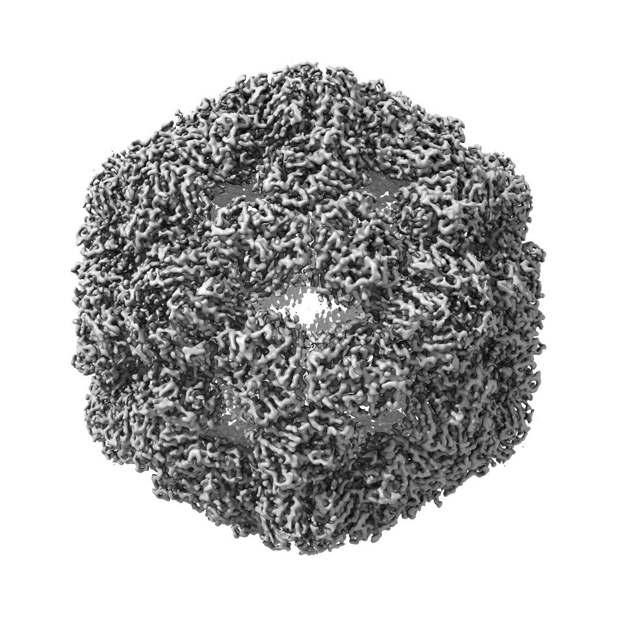
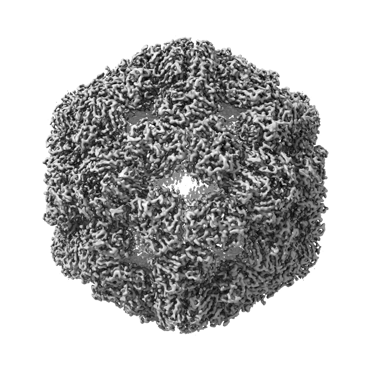
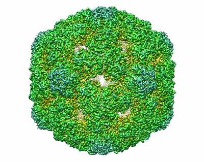
The structure of Enterovirus D68 procapsid
EMD-9632
Single-particle3.8 Å
 Deposition: 26/08/2018
Map released: 07/11/2018
Last modified: 23/03/2022
Buffer
pH: 7.4
Vitrification
Cryogen name: ETHANE
Microscope: FEI TECNAI F30
Illumination mode: FLOOD BEAM
Imaging mode: BRIGHT FIELD
Electron source: FIELD EMISSION GUN
Acceleration voltage: 300 kV
Illumination mode: FLOOD BEAM
Imaging mode: BRIGHT FIELD
Electron source: FIELD EMISSION GUN
Acceleration voltage: 300 kV
Image Recording
[1]
Final
reconstruction
Resolution: 3.8
Å
(
BY AUTHOR)
Resolution method: FSC 0.143 CUT-OFF
Number of images used: 13849
Resolution method: FSC 0.143 CUT-OFF
Number of images used: 13849
⌯ Applied Symmetry
Point group:
I
Software
[1]
| Name | Version | Details |
|---|---|---|
| RELION | 2.0 | - |
⦨ Initial angle
assignment
Type:
MAXIMUM LIKELIHOOD
⦩ Final angle assignment
Type:
MAXIMUM LIKELIHOOD
Format: CCP4
Data type: IMAGE STORED AS FLOATING POINT NUMBER (4 BYTES)
Annotation details: None
Data type: IMAGE STORED AS FLOATING POINT NUMBER (4 BYTES)
Annotation details: None
⬡ Geometry
| X | Y | Z | |
|---|---|---|---|
| Dimensions | 450 | 450 | 450 |
| Origin | 0 | 0 | 0 |
| Spacing | 450 | 450 | 450 |
| Voxel size | 1.128 Å | 1.128 Å | 1.128 Å |
Contour list
| Primary | Level | Source |
|---|---|---|
| True | 0.0613 | AUTHOR |