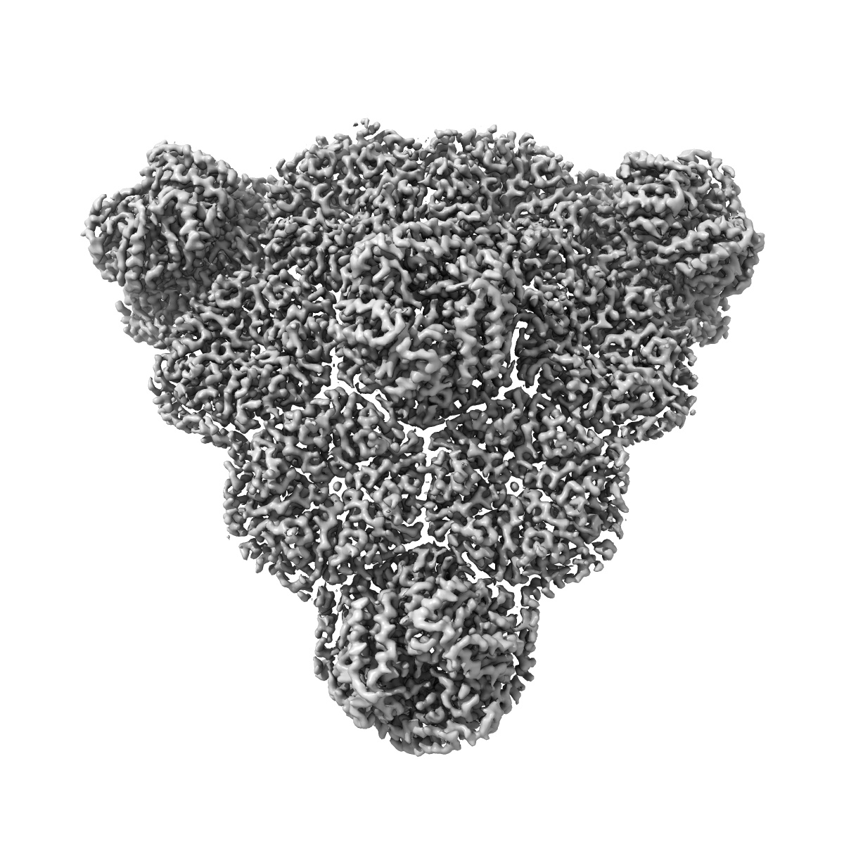
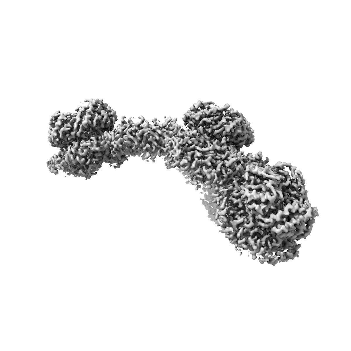
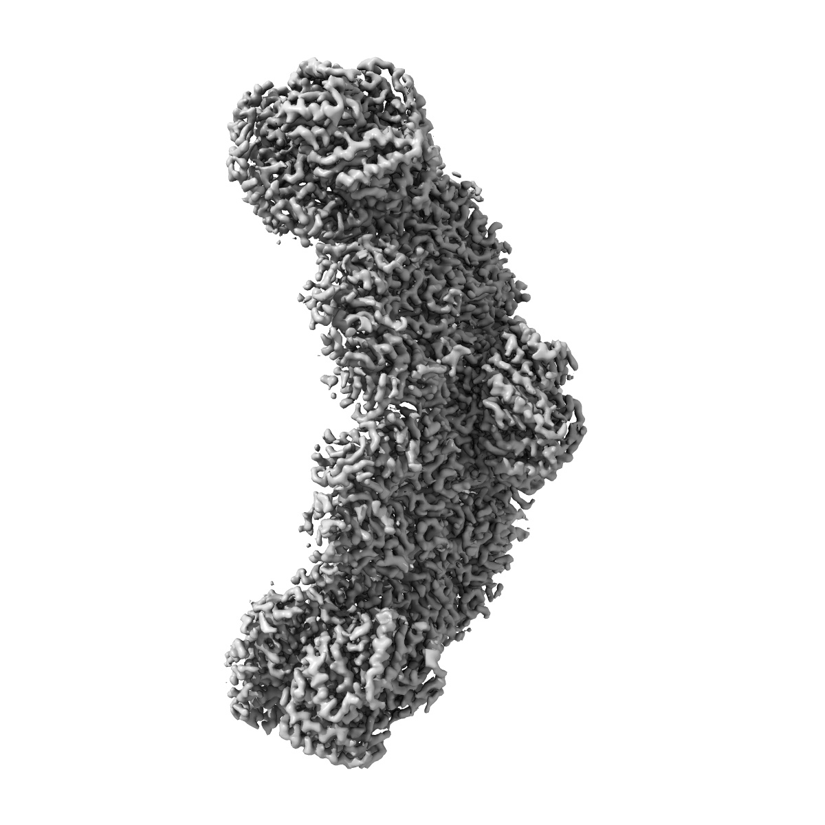
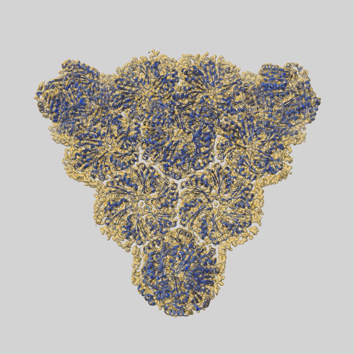
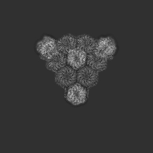
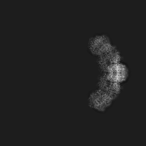
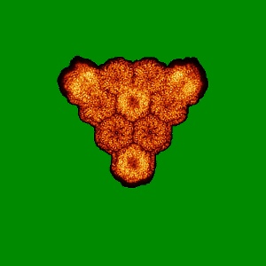
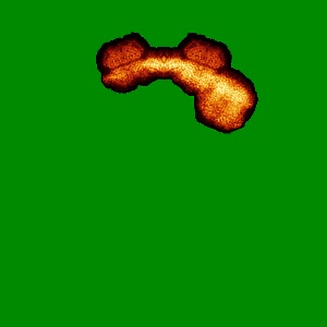
EMD-9313
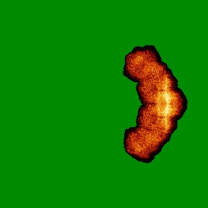
Cryo-EM structure of the HO BMC shell: subregion classified for BMC-T: TD-TDTDTD
EMD-9313
Single-particle3.5 Å
 Deposition: 06/11/2018
Map released: 13/03/2019
Last modified: 13/03/2024
Concentration: 3
mg/mL
Buffer
pH: 7.4
Buffer components [3]:
Buffer components [3]:
| Name | Formula | Concentration | ChEBI |
|---|---|---|---|
| Tris-HCl | - | 20.0 mM | - |
| Sodium chloride | NaCl | 50.0 mM | |
| NP-40 substitute | - | 0.01 % | - |
Grid
Details: Unspecified
Support Film [2]
| Material | Topology | Thickness |
|---|---|---|
| CARBON | HOLEY | - |
| CARBON | CONTINUOUS | - |
Vitrification
Cryogen name: ETHANE
Chamber humidity: 100%
Chamber temperature: 277 K
Instrument: FEI VITROBOT MARK IV
Details: 5-7 second incubation of the sample on the grid before blotting and plunging.
Chamber humidity: 100%
Chamber temperature: 277 K
Instrument: FEI VITROBOT MARK IV
Details: 5-7 second incubation of the sample on the grid before blotting and plunging.
Microscope: FEI TITAN
Illumination mode: FLOOD BEAM
Imaging mode: BRIGHT FIELD
Electron source: FIELD EMISSION GUN
Acceleration voltage: 300 kV
C2 aperture diameter: 50.0 µm
Nominal CS: 2.7 mm
Calibrated defocus: 1.0 µm - 3.5 µm
Calibrated magnification: 48543.0
Specimen holder model: GATAN 626 SINGLE TILT LIQUID NITROGEN CRYO TRANSFER HOLDER
Cooling holder cryogen: NITROGEN
Alignment procedure: COMA FREE
Image Recording
[1]
Detector model:
GATAN K2 SUMMIT (4k x 4k)
Detector mode: COUNTING
Dimensions: 3838 pixel x 3710 pixel
Frames per image: 1-30
Number of grids: 1
Number of real images: 928
Average exposure time: 4.5 s
Average electron dose per image: 25.0 e/Å2
Details: 928 images retained after inspection for image quality.
Detector mode: COUNTING
Dimensions: 3838 pixel x 3710 pixel
Frames per image: 1-30
Number of grids: 1
Number of real images: 928
Average exposure time: 4.5 s
Average electron dose per image: 25.0 e/Å2
Details: 928 images retained after inspection for image quality.
Final
reconstruction
Resolution: 3.5
Å
(
BY AUTHOR)
Resolution method: FSC 0.143 CUT-OFF
Number of images used: 106640
Algorithm: FOURIER SPACE
Resolution method: FSC 0.143 CUT-OFF
Number of images used: 106640
Algorithm: FOURIER SPACE
⌯ Applied Symmetry
Point group:
C1
Software
[1]
| Name | Version | Details |
|---|---|---|
| RELION | 1.4 | - |
Startup model
[1]
⦨ Initial angle
assignment
⦩ Final angle assignment
Particle selection
[1]
| Selected | Ref. model | Method | Software | Details |
|---|---|---|---|---|
| 31800 | - | - | - | 1000 particles were picked manually to generate reference templates for subsequent auto-picking in RELION 1.4. |
Final 3D classification
Number of classes:
2
Details: Three sequential classifications for four BMC-T positions in total, using the symmetry-expanded particle dataset
Details: Three sequential classifications for four BMC-T positions in total, using the symmetry-expanded particle dataset
Software
[1]
| Name | Version | Details |
|---|---|---|
| RELION | 1.4 | - |
Format: CCP4
Data type: IMAGE STORED AS FLOATING POINT NUMBER (4 BYTES)
Annotation details: Four BMC-T positions classified: TD-TDTDTD
Data type: IMAGE STORED AS FLOATING POINT NUMBER (4 BYTES)
Annotation details: Four BMC-T positions classified: TD-TDTDTD
⬡ Geometry
| X | Y | Z | |
|---|---|---|---|
| Dimensions | 512 | 512 | 512 |
| Origin | 0 | 0 | 0 |
| Spacing | 512 | 512 | 512 |
| Voxel size | 1.03 Å | 1.03 Å | 1.03 Å |
Contour list
| Primary | Level | Source |
|---|---|---|
| True | 0.03 | AUTHOR |