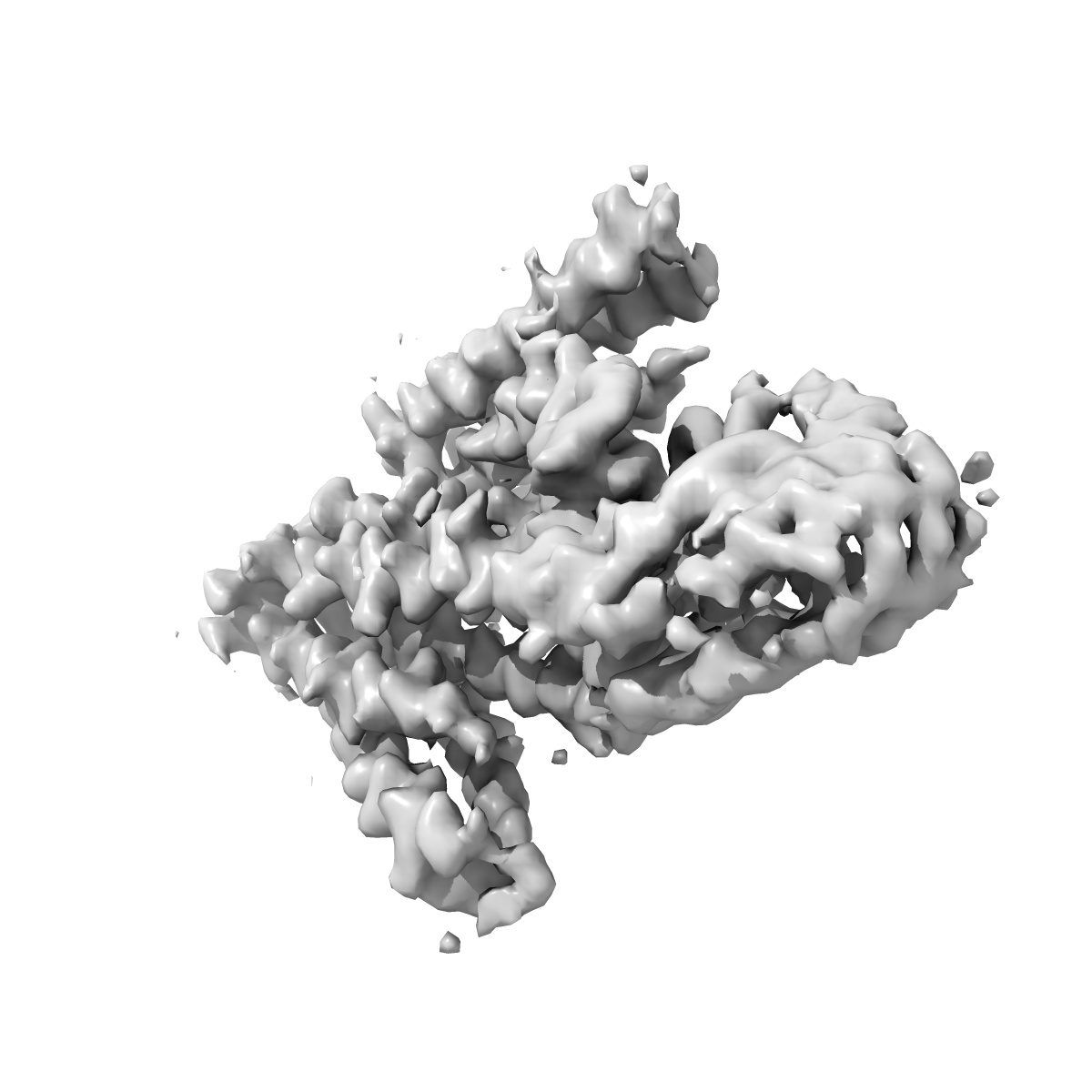
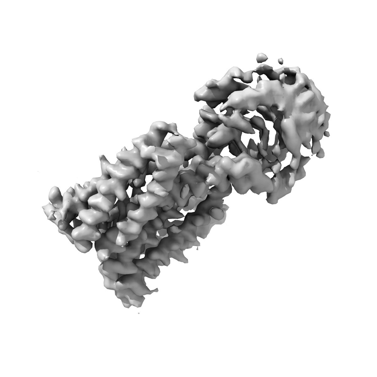
EMD-8728
CryoEM Structure of the Zinc Transporter YiiP from helical crystals
EMD-8728
Helical reconstruction4.1 Å
 Deposition: 10/05/2017
Map released: 14/03/2018
Last modified: 13/03/2024
Concentration: 0.6
mg/mL
Details: The protein was purified in 0.2% n-dodecyl beta-D-maltoside
Details: The protein was purified in 0.2% n-dodecyl beta-D-maltoside
Buffer
pH: 7.0
Buffer components [3]:
Details: Buffer was changed twice per day.
Buffer components [3]:
| Name | Formula | Concentration | ChEBI |
|---|---|---|---|
| Sodium chloride | NaCl | 100.0 mM | |
| Magnesium chloride | MgCl2 | 5.0 mM | |
| Sodium azide | NaN3 | 5.0 mM |
Grid
Vitrification
Microscope: FEI TITAN KRIOS
Illumination mode: FLOOD BEAM
Imaging mode: BRIGHT FIELD
Electron source: LAB6
Acceleration voltage: 300 kV
Nominal CS: 2.7 mm
Nominal defocus: 1.2 µm - 2.5 µm
Calibrated defocus: 1.0 µm - 5.4 µm
Nominal magnification: 22500.0
Specimen holder model: FEI TITAN KRIOS AUTOGRID HOLDER
Cooling holder cryogen: NITROGEN
Alignment procedure: COMA FREE
Image Recording
[1]
Detector model:
GATAN K2 SUMMIT (4k x 4k)
Detector mode: COUNTING
Dimensions: 3710 pixel x 3838 pixel
Frames per image: 2-16
Number of grids: 3
Number of real images: 2743
Average exposure time: 10.0 s
Average electron dose per image: 70.0 e/Å2
Detector mode: COUNTING
Dimensions: 3710 pixel x 3838 pixel
Frames per image: 2-16
Number of grids: 3
Number of real images: 2743
Average exposure time: 10.0 s
Average electron dose per image: 70.0 e/Å2
Final
reconstruction
Resolution: 4.1
Å
(
BY AUTHOR)
Resolution method: FSC 0.143 CUT-OFF
Number of images used: 72333
Resolution method: FSC 0.143 CUT-OFF
Number of images used: 72333
⌯ Applied Symmetry
Software
[1]
| Name | Version | Details |
|---|---|---|
| RELION | 2.0 | - |
Startup model
[1]
Type:
OTHER
Details:A map was obtained previously with images acquired on a JEOL2100 microscope. This map was used as a startup model (That map was obtained as follow: we started with a noisy cylinder. Then, a first reconstruction was obtained with Helicon (from SPARX) and refined with Frealix).
Details:A map was obtained previously with images acquired on a JEOL2100 microscope. This map was used as a startup model (That map was obtained as follow: we started with a noisy cylinder. Then, a first reconstruction was obtained with Helicon (from SPARX) and refined with Frealix).
⦩ Final angle assignment
Segment selection
[1]
| Number selected | Segment length | Segment overlap | Total filament length | Details |
|---|---|---|---|---|
| 141904 | - | - | - | 2293 filaments selected with SPARX/EMAN2, then windowed into 141,904 segments (450x450 pixel overlapping segments) |
Format: CCP4
Data type: IMAGE STORED AS FLOATING POINT NUMBER (4 BYTES)
Annotation details: Primary map
Data type: IMAGE STORED AS FLOATING POINT NUMBER (4 BYTES)
Annotation details: Primary map
⬡ Geometry
| X | Y | Z | |
|---|---|---|---|
| Dimensions | 131 | 131 | 131 |
| Origin | 0 | 0 | 0 |
| Spacing | 131 | 131 | 131 |
| Voxel size | 1.07 Å | 1.07 Å | 1.07 Å |
Contour list
| Primary | Level | Source |
|---|---|---|
| True | 0.018 | AUTHOR |