EMD-7442
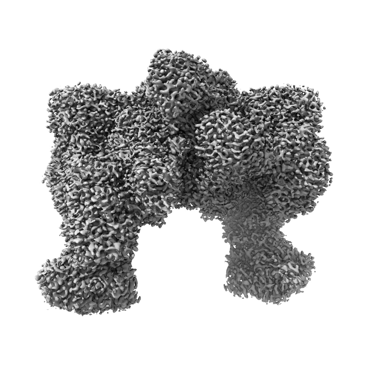
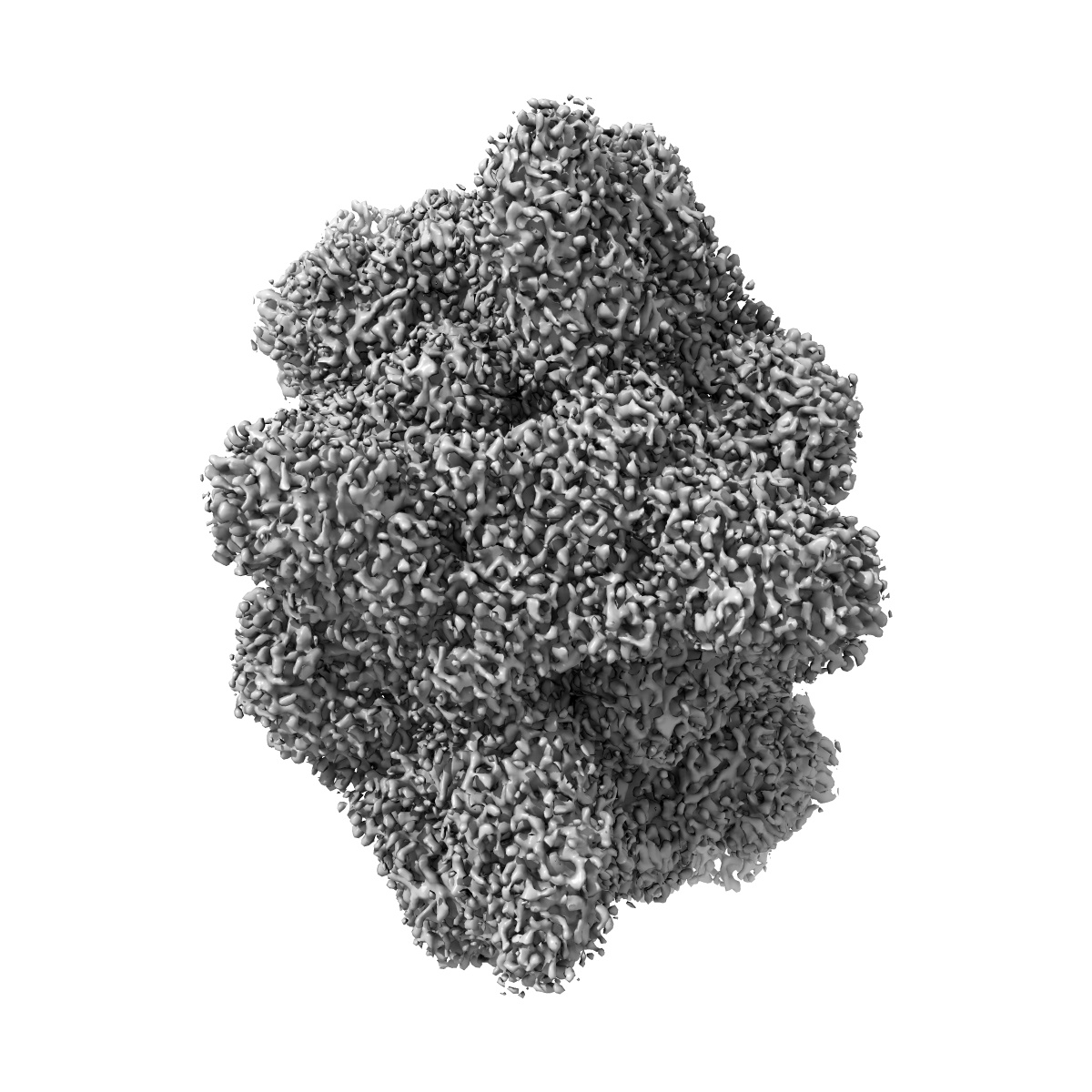
Electron cryo-microscopy of the eukaryotic translation initiation factor 2B from Homo sapiens
EMD-7442
Single-particle2.8 Å
 Deposition: 31/01/2018
Map released: 11/04/2018
Last modified: 25/11/2020
Buffer
pH: 7.5
Vitrification
Cryogen name: ETHANE
Chamber humidity: 100%
Chamber temperature: 277 K
Instrument: FEI VITROBOT MARK IV
Chamber humidity: 100%
Chamber temperature: 277 K
Instrument: FEI VITROBOT MARK IV
Microscope: FEI TITAN KRIOS
Illumination mode: FLOOD BEAM
Imaging mode: BRIGHT FIELD
Electron source: FIELD EMISSION GUN
Acceleration voltage: 300 kV
Illumination mode: FLOOD BEAM
Imaging mode: BRIGHT FIELD
Electron source: FIELD EMISSION GUN
Acceleration voltage: 300 kV
Image Recording
[1]
Detector model:
GATAN K2 SUMMIT (4k x 4k)
Detector mode: SUPER-RESOLUTION
Average electron dose per image: 1.63 e/Å2
Detector mode: SUPER-RESOLUTION
Average electron dose per image: 1.63 e/Å2
Final
reconstruction
Resolution: 2.8
Å
(
BY AUTHOR)
Resolution method: FSC 0.5 CUT-OFF
Number of images used: 202125
Resolution method: FSC 0.5 CUT-OFF
Number of images used: 202125
⌯ Applied Symmetry
Point group:
C2
Software
[1]
| Name | Version | Details |
|---|---|---|
| FREALIGN | - | - |
Startup model
[1]
⦨ Initial angle
assignment
⦩ Final angle assignment
Particle selection
[1]
| Selected | Ref. model | Method | Software | Details |
|---|---|---|---|---|
| 202125 | - | - | - | - |
CTF correction
Software
[1]
| Name | Version | Details |
|---|---|---|
| Gctf | 1.06 | - |
Format: CCP4
Data type: IMAGE STORED AS FLOATING POINT NUMBER (4 BYTES)
Annotation details: Primary
Data type: IMAGE STORED AS FLOATING POINT NUMBER (4 BYTES)
Annotation details: Primary
⬡ Geometry
| X | Y | Z | |
|---|---|---|---|
| Dimensions | 458 | 458 | 458 |
| Origin | 0 | 0 | 0 |
| Spacing | 458 | 458 | 458 |
| Voxel size | 0.83799994 Å | 0.83799994 Å | 0.83799994 Å |
Contour list
| Primary | Level | Source |
|---|---|---|
| True | 4.62 | AUTHOR |