EMD-7119
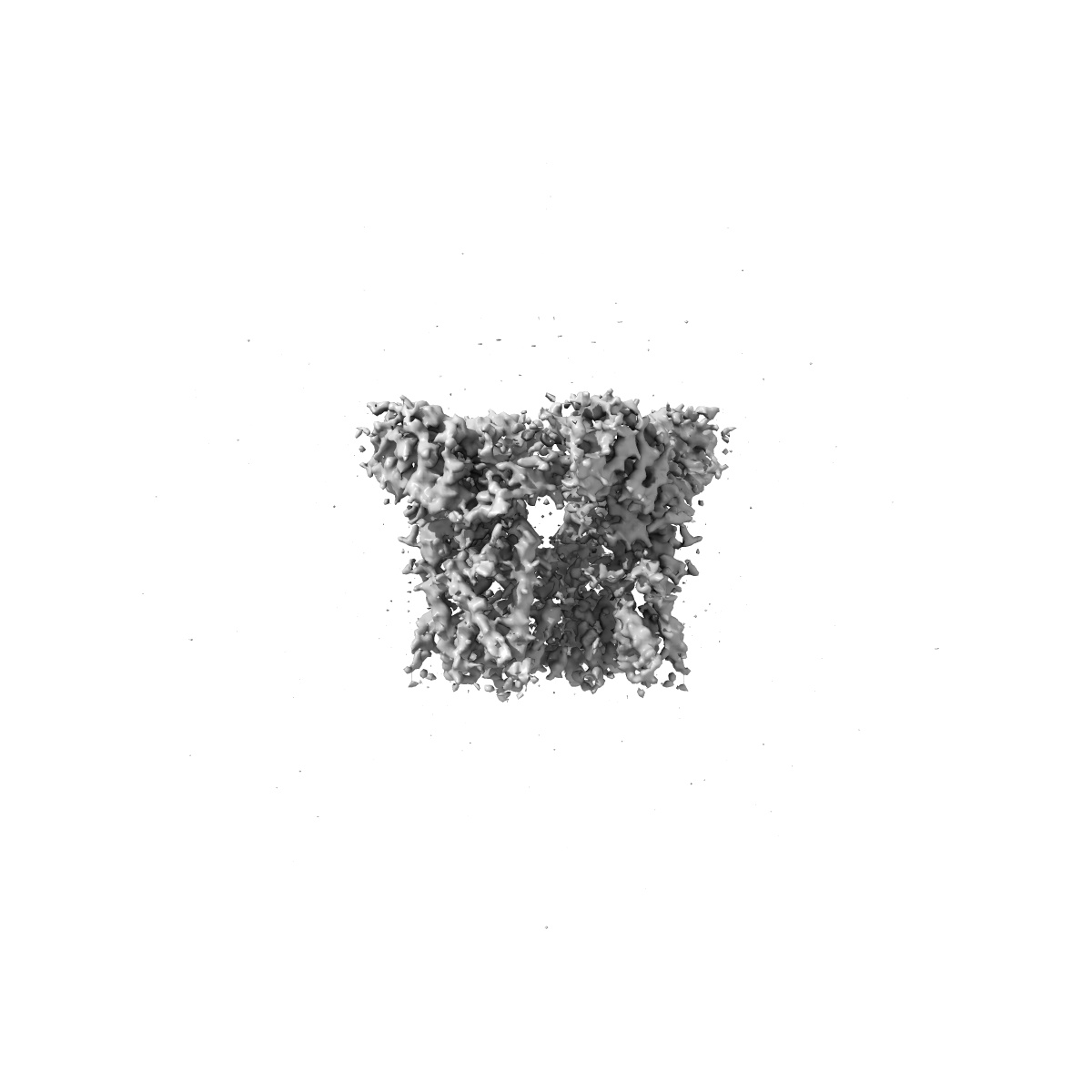
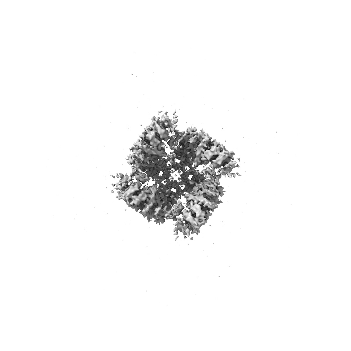
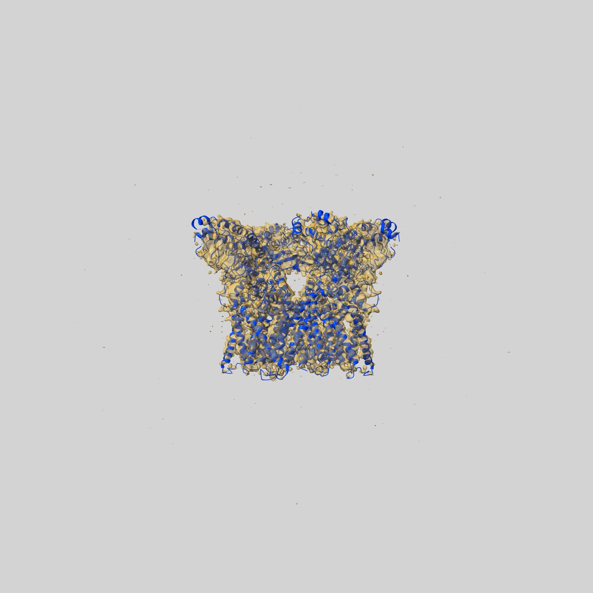
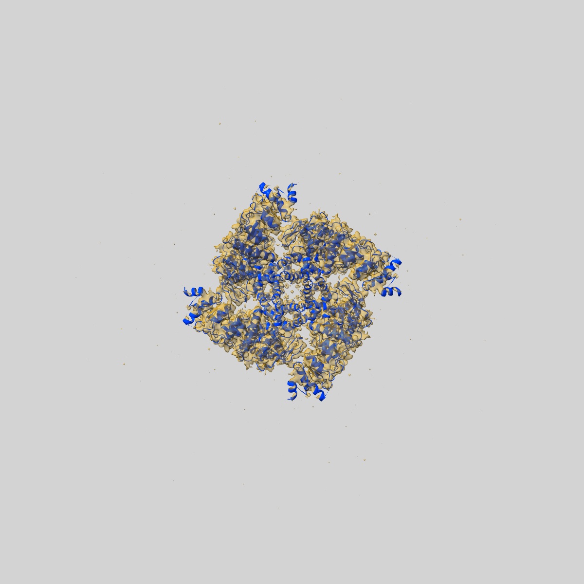
TRPV2 ion channel in partially closed state
EMD-7119
Single-particle3.6 Å
 Deposition: 18/11/2017
Map released: 05/12/2018
Last modified: 13/03/2024
Concentration: .5
mg/mL
Buffer
pH: 8.0
Grid
Details: Unspecified
Vitrification
Microscope: JEOL 3200FSC
Illumination mode: SPOT SCAN
Imaging mode: BRIGHT FIELD
Electron source: FIELD EMISSION GUN
Acceleration voltage: 300 kV
Illumination mode: SPOT SCAN
Imaging mode: BRIGHT FIELD
Electron source: FIELD EMISSION GUN
Acceleration voltage: 300 kV
Image Recording
[1]
Final
reconstruction
Resolution: 3.6
Å
(
BY AUTHOR)
Resolution method: FSC 0.143 CUT-OFF
Number of images used: 50509
Resolution method: FSC 0.143 CUT-OFF
Number of images used: 50509
⌯ Applied Symmetry
Point group:
C4
Software
[1]
| Name | Version | Details |
|---|---|---|
| RELION | 1.4 | - |
Startup model
[1]
Type:
NONE
⦨ Initial angle
assignment
Type:
RANDOM ASSIGNMENT
⦩ Final angle assignment
Particle selection
[1]
| Selected | Ref. model | Method | Software | Details |
|---|---|---|---|---|
| 160100 | - | - | - | - |
Format: CCP4
Data type: IMAGE STORED AS FLOATING POINT NUMBER (4 BYTES)
Annotation details: TRPV2 ion channel in a partially closed state and with a deletion in the pore turret domain.
Data type: IMAGE STORED AS FLOATING POINT NUMBER (4 BYTES)
Annotation details: TRPV2 ion channel in a partially closed state and with a deletion in the pore turret domain.
⬡ Geometry
| X | Y | Z | |
|---|---|---|---|
| Dimensions | 220 | 220 | 220 |
| Origin | 0 | 0 | 0 |
| Spacing | 220 | 220 | 220 |
| Voxel size | 1.23 Å | 1.23 Å | 1.23 Å |
Contour list
| Primary | Level | Source |
|---|---|---|
| True | 0.018 | AUTHOR |