EMD-6976
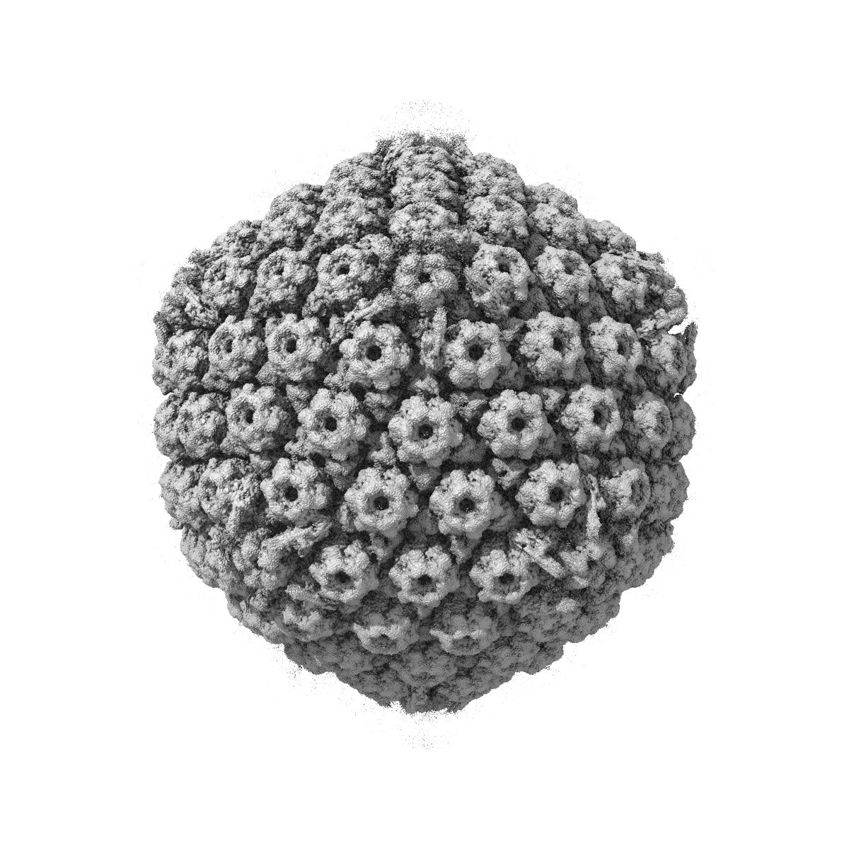
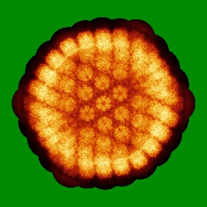
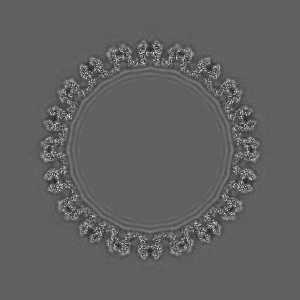
Structure of the Herpes simplex virus type 2 C-capsid with capsid-vertex-specific component
EMD-6976
Single-particle3.75 Å
 Deposition: 31/05/2018
Map released: 10/10/2018
Last modified: 06/11/2019
Buffer
pH: 7.4
Vitrification
Cryogen name: ETHANE
Microscope: FEI TITAN KRIOS
Illumination mode: FLOOD BEAM
Imaging mode: OTHER
Electron source: FIELD EMISSION GUN
Acceleration voltage: 300 kV
Illumination mode: FLOOD BEAM
Imaging mode: OTHER
Electron source: FIELD EMISSION GUN
Acceleration voltage: 300 kV
Image Recording
[1]
Final
reconstruction
Resolution: 3.75
Å
(
BY AUTHOR)
Resolution method: FSC 0.143 CUT-OFF
Number of images used: 56901
Resolution method: FSC 0.143 CUT-OFF
Number of images used: 56901
⌯ Applied Symmetry
Point group:
C1
⦨ Initial angle
assignment
Type:
MAXIMUM LIKELIHOOD
⦩ Final angle assignment
Type:
MAXIMUM LIKELIHOOD
Format: CCP4
Data type: IMAGE STORED AS FLOATING POINT NUMBER (4 BYTES)
Annotation details: Full_map
Data type: IMAGE STORED AS FLOATING POINT NUMBER (4 BYTES)
Annotation details: Full_map
⬡ Geometry
| X | Y | Z | |
|---|---|---|---|
| Dimensions | 1200 | 1200 | 1200 |
| Origin | 0 | 0 | 0 |
| Spacing | 1200 | 1200 | 1200 |
| Voxel size | 1.38 Å | 1.38 Å | 1.38 Å |
Contour list
| Primary | Level | Source |
|---|---|---|
| True | 1.0 | AUTHOR |