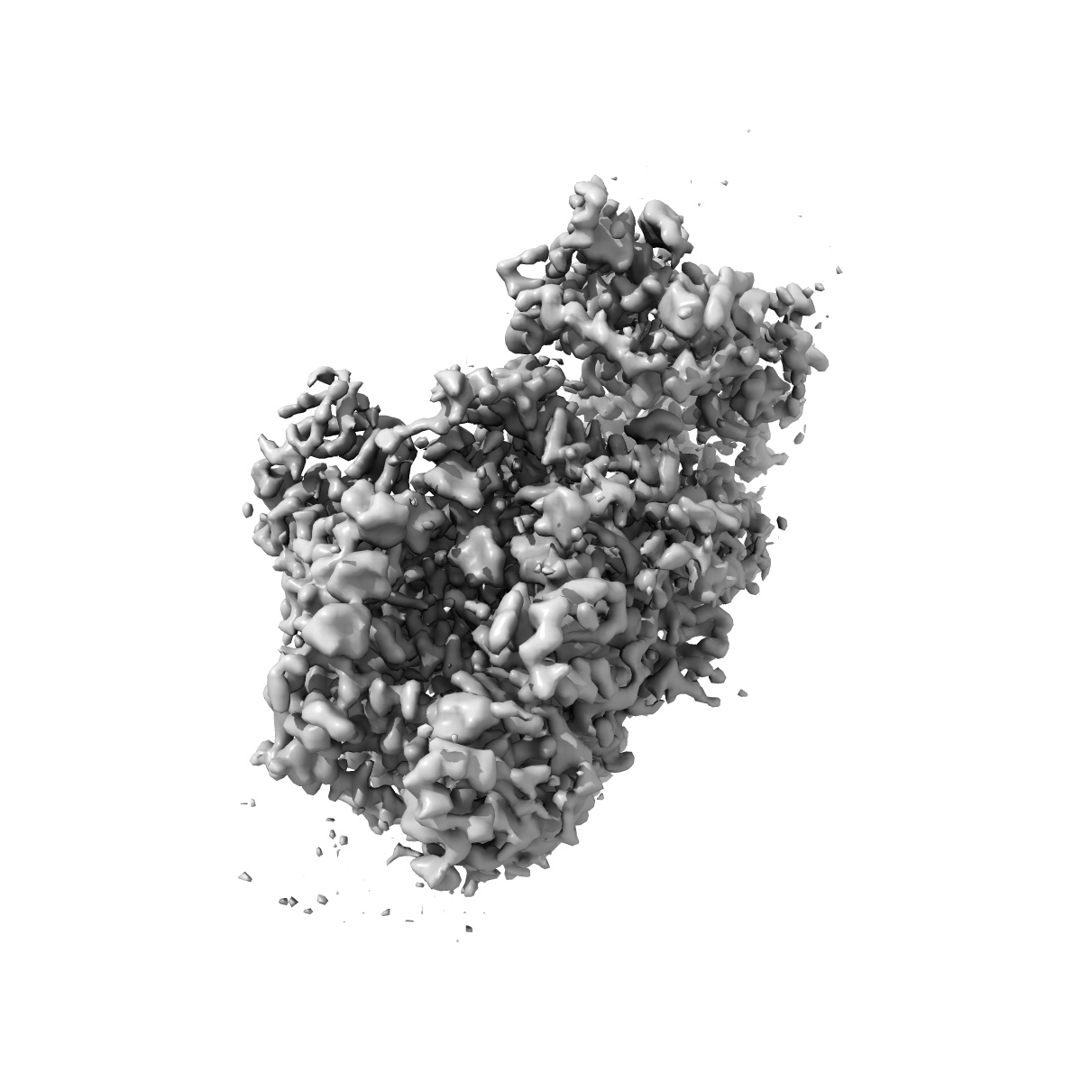
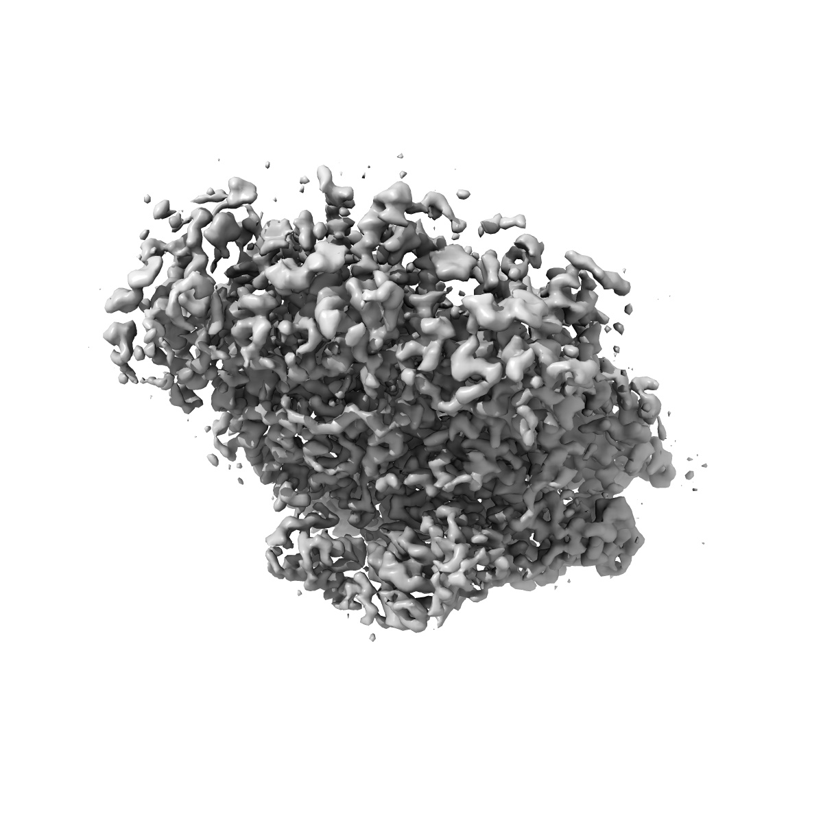
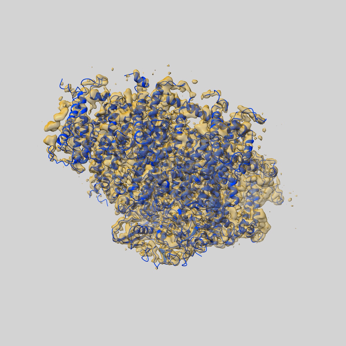
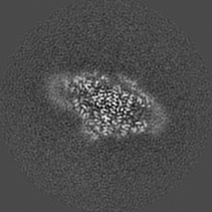
EMD-6930
Cryo-EM structure of the red algal PSI-LHCR
EMD-6930
Single-particle3.63 Å
 Deposition: 08/03/2018
Map released: 11/04/2018
Last modified: 22/01/2020
Microscope: FEI TITAN KRIOS
Illumination mode: FLOOD BEAM
Imaging mode: BRIGHT FIELD
Electron source: FIELD EMISSION GUN
Acceleration voltage: 300 kV
Illumination mode: FLOOD BEAM
Imaging mode: BRIGHT FIELD
Electron source: FIELD EMISSION GUN
Acceleration voltage: 300 kV
Image Recording
[1]
Format: CCP4
Data type: IMAGE STORED AS FLOATING POINT NUMBER (4 BYTES)
Data type: IMAGE STORED AS FLOATING POINT NUMBER (4 BYTES)
⬡ Geometry
| X | Y | Z | |
|---|---|---|---|
| Dimensions | 280 | 280 | 280 |
| Origin | 0 | 0 | 0 |
| Spacing | 280 | 280 | 280 |
| Voxel size | 1.05 Å | 1.05 Å | 1.05 Å |
Contour list
| Primary | Level | Source |
|---|---|---|
| True | 0.087 | AUTHOR |