EMD-5540
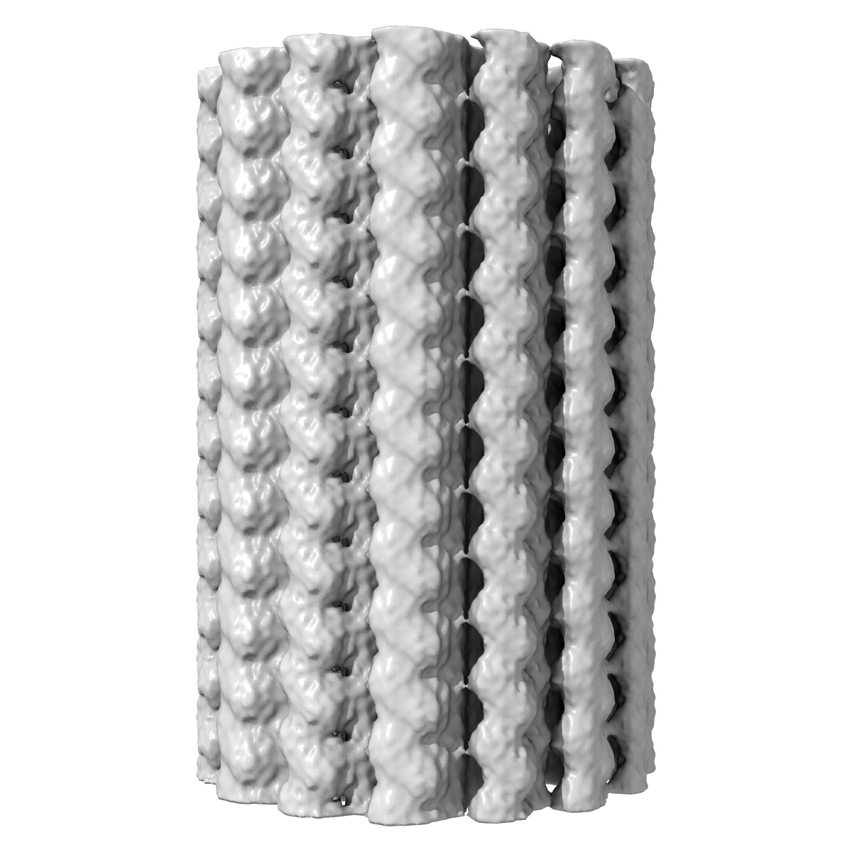
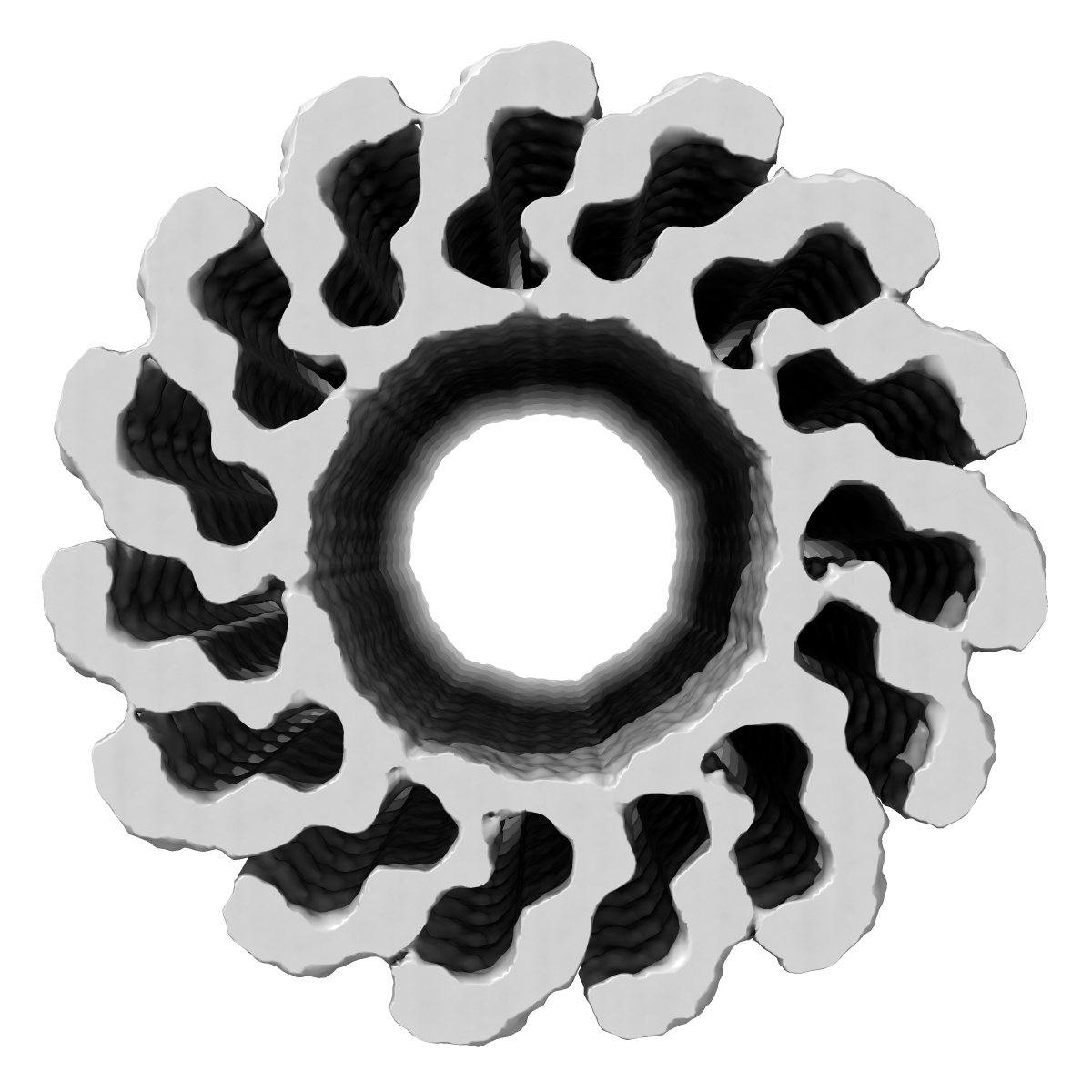
3D membrane-bound structure of FVIII bound to single lipid bilayer nanotubes
EMD-5540
Helical reconstruction15.0 Å
 Deposition: 12/12/2012
Map released: 28/08/2013
Last modified: 28/08/2013
Concentration: 1
mg/mL
Details: The protein was mixed in 1:1 w/w ratio with lipid nanotubes solution
Details: The protein was mixed in 1:1 w/w ratio with lipid nanotubes solution
Buffer
pH: 7.4
Details: 20 mM Tris-HCl 150 mM NaCl, 20 mM EDTA
Details: 20 mM Tris-HCl 150 mM NaCl, 20 mM EDTA
Grid
Details: 300 mesh R2x2 Quantifoil grids
Vitrification
Cryogen name: ETHANE
Chamber humidity: 100%
Chamber temperature: 106 K
Instrument: FEI VITROBOT MARK III
Method: Blot for 4.5 seconds before plunging
Chamber humidity: 100%
Chamber temperature: 106 K
Instrument: FEI VITROBOT MARK III
Method: Blot for 4.5 seconds before plunging
Microscope: JEOL 2010F
Illumination mode: FLOOD BEAM
Imaging mode: BRIGHT FIELD
Electron source: FIELD EMISSION GUN
Acceleration voltage: 200 kV
Nominal CS: 2.0 mm
Nominal defocus: -0.7 µm - -4.4 µm
Nominal magnification: 52000.0
Calibrated magnification: 52000.0
Specimen holder model: GATAN LIQUID NITROGEN
Alignment procedure: LEGACY (Astigmatism: corrected at 400,000 times magnification, Electron beam tilt params: )
Temperature
Minimum: 90
K
Average: 99 K
Maximum: 100 K
Average: 99 K
Maximum: 100 K
Image Recording
[1]
Detector category:
CCD
Detector model: GATAN ULTRASCAN 4000 (4k x 4k)
Sampling interval: 15 µm
Number of real images: 69
Average electron dose per image: 16 e/Å2
Details: Each image was acquired for 1 second.
Detector model: GATAN ULTRASCAN 4000 (4k x 4k)
Sampling interval: 15 µm
Number of real images: 69
Average electron dose per image: 16 e/Å2
Details: Each image was acquired for 1 second.
Details: The 2D analysis was performed with EMAN2 and the helical reconstruction with the IHRSR algorithm
Final
reconstruction
Resolution: 15.0
Å
(
BY AUTHOR)
Resolution method: OTHER
Algorithm: OTHER
Details: The final 3D reconstructions was calculated from a set of 2043 helical segments cut off from the selected helical tubes at 256 x 256 pixels with 10% overlap.
Resolution method: OTHER
Algorithm: OTHER
Details: The final 3D reconstructions was calculated from a set of 2043 helical segments cut off from the selected helical tubes at 256 x 256 pixels with 10% overlap.
⌯ Applied Symmetry
ΔΖ:
7.6
Å
ΔΦ: 0.5°
ΔΦ: 0.5°
Software
[1]
| Name | Version | Details |
|---|---|---|
| EMAN2, IHRSR | - | - |
CTF correction
Details:EMAN2, only phase correction
Format: CCP4
Data type: IMAGE STORED AS FLOATING POINT NUMBER (4 BYTES)
Annotation details: Helical reconstruction of membrane-bound Factor vIII light chain bound to single bilayer lipid nanotubes
Details: ::::EMDATABANK.org::::EMD-5540::::
Data type: IMAGE STORED AS FLOATING POINT NUMBER (4 BYTES)
Annotation details: Helical reconstruction of membrane-bound Factor vIII light chain bound to single bilayer lipid nanotubes
Details: ::::EMDATABANK.org::::EMD-5540::::
⬡ Geometry
| X | Y | Z | |
|---|---|---|---|
| Dimensions | 256 | 256 | 256 |
| Origin | 0 | 0 | 0 |
| Spacing | 256 | 256 | 256 |
| Voxel size | 3.0 Å | 3.0 Å | 3.0 Å |
Contour list
| Primary | Level | Source |
|---|---|---|
| True | 0.006 | AUTHOR |