EMD-5188
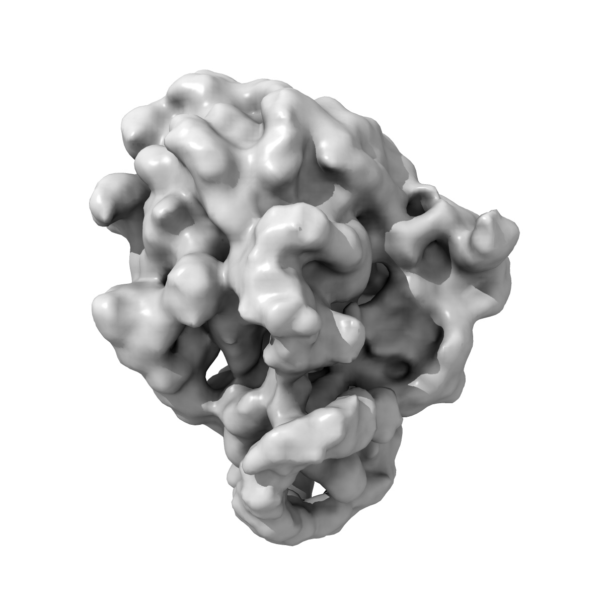
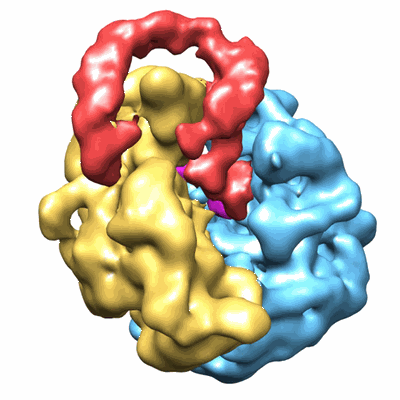
tmRNA-SmpB: a journey to the center of the bacterial ribosome
EMD-5188
Single-particle13.0 Å
 Deposition: 16/04/2010
Map released: 13/10/2010
Last modified: 06/03/2013
Buffer
pH: 7.5
Details: 5mM Hepes-KOH (pH 7.5), 10mM NH4Cl, 10mM MgOAc, 50mM KCl, 0.1mM EDTA and 6mM BetaME
Details: 5mM Hepes-KOH (pH 7.5), 10mM NH4Cl, 10mM MgOAc, 50mM KCl, 0.1mM EDTA and 6mM BetaME
Grid
Details: Quantifoil holey-carbon grids previously glow-discharged
Microscope: JEOL 2200FS
Illumination mode: FLOOD BEAM
Imaging mode: BRIGHT FIELD
Electron source: FIELD EMISSION GUN
Acceleration voltage: 200 kV
Nominal CS: 2 mm
Nominal defocus: 0.9 µm - 2.2 µm
Nominal magnification: 50000.0
Calibrated magnification: 45700.0
Specimen holder model: GATAN LIQUID NITROGEN
Specimen holder details: Eucentric
Temperature
Average: 95
K
Specialist optics
Energy filter
Image Recording
[1]
Detector category:
FILM
Detector model: KODAK SO-163 FILM
Scanner: NIKON SUPER COOLSCAN 9000
Sampling interval: 7.5 µm
Number of real images: 70
Bits per pixel: 8.0
Detector model: KODAK SO-163 FILM
Scanner: NIKON SUPER COOLSCAN 9000
Sampling interval: 7.5 µm
Number of real images: 70
Bits per pixel: 8.0
Final
reconstruction
Resolution: 13.0
Å
(
BY AUTHOR)
Resolution method: FSC 0.5 CUT-OFF
Number of images used: 49061
Resolution method: FSC 0.5 CUT-OFF
Number of images used: 49061
⌯ Applied Symmetry
Point group:
C1
Software
[1]
| Name | Version | Details |
|---|---|---|
| IMAGIC-V | - | - |
CTF correction
Details:Each particle
Format: CCP4
Data type: IMAGE STORED AS FLOATING POINT NUMBER (4 BYTES)
Annotation details: Structure of tmRNA-SMPB complex accomodated into a stalled bacterial ribosome
Details: ::::EMDATABANK.org::::EMD-5188::::
Data type: IMAGE STORED AS FLOATING POINT NUMBER (4 BYTES)
Annotation details: Structure of tmRNA-SMPB complex accomodated into a stalled bacterial ribosome
Details: ::::EMDATABANK.org::::EMD-5188::::
⬡ Geometry
| X | Y | Z | |
|---|---|---|---|
| Dimensions | 128 | 128 | 128 |
| Origin | -64 | -64 | -64 |
| Spacing | 128 | 128 | 128 |
| Voxel size | 3.28 Å | 3.28 Å | 3.28 Å |
Contour list
| Primary | Level | Source |
|---|---|---|
| True | 0.01 | AUTHOR |