EMD-5006
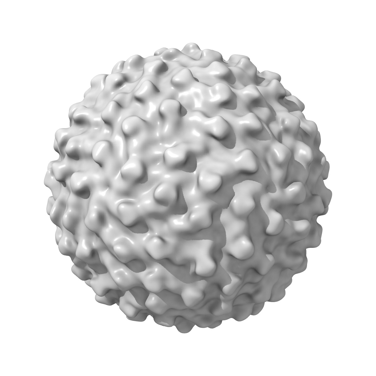
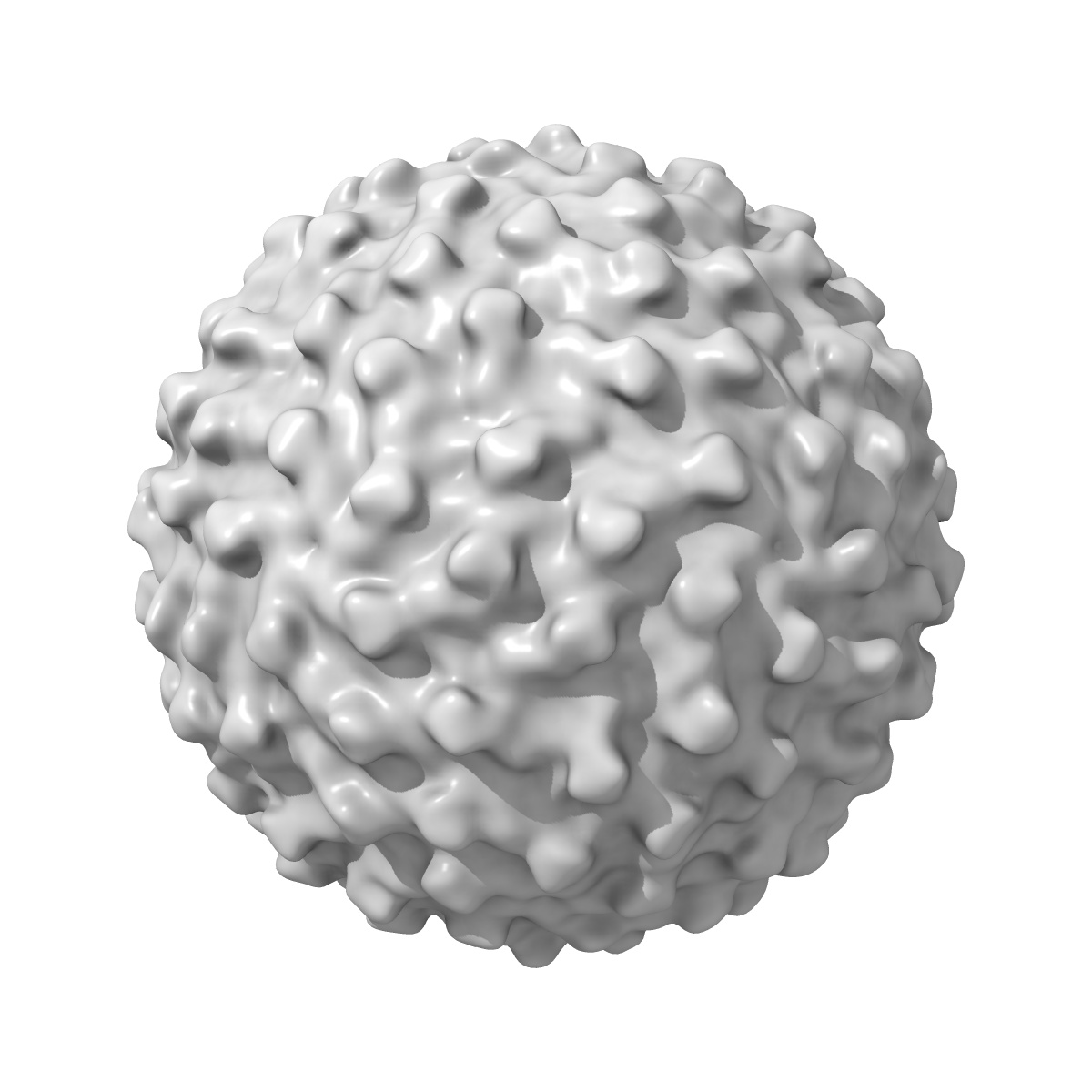
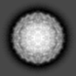
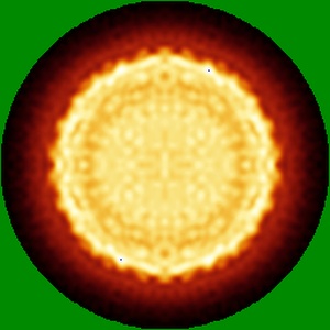
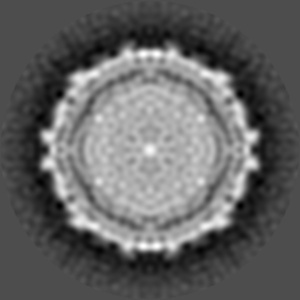
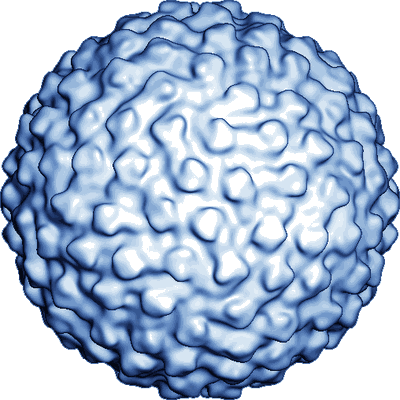
Structure of immature Dengue virus at low pH
EMD-5006
Single-particle25.0 Å
 Deposition: 26/02/2008
Map released: 22/04/2009
Last modified: 08/07/2011
Buffer
pH: 6.0
Details: The virus was mixed in NTE buffer (10 mM Tris, 120 mM NaCl, and 1 mM EDTA at pH 8) with 50 mM MES, 120 mM NaCl at pH 5.6 to yield a final pH of 6
Details: The virus was mixed in NTE buffer (10 mM Tris, 120 mM NaCl, and 1 mM EDTA at pH 8) with 50 mM MES, 120 mM NaCl at pH 5.6 to yield a final pH of 6
Grid
Details: 400 mesh copper grid
Vitrification
Cryogen name: ETHANE
Instrument: OTHER
Method: A small vial of ethane is placed inside a larger liquid nitrogen reservoir. The grid holding a few microliters of the sample is held in place at the bottom of a plunger by the means of fine tweezers. Once the ethane in the vial is completely frozen, it needs to be slightly melted. When the liquid ethane is ready, a piece of filter paper is then pressed against the sample to blot of excess buffer, sufficient to leave a thin layer on the grid. After a predetermined time, the filter paper is removed, and the plunger is allowed to drop into the liquid ethane. Once the grid enters the liquid ethane, the sample is rapidly frozen, and the grid is transferred under liquid nitrogen to a storage box immersed liquid nitrogen for later use in the microscope.
Microscope: FEI/PHILIPS CM200FEG
Illumination mode: FLOOD BEAM
Imaging mode: BRIGHT FIELD
Electron source: FIELD EMISSION GUN
Acceleration voltage: 200 kV
Nominal CS: 2.0 mm
Nominal defocus: 1.4 µm - 2.9 µm
Nominal magnification: 50000.0
Calibrated magnification: 51040.0
Specimen holder model: GATAN LIQUID NITROGEN
Specimen holder details: Eucentric
Minimum tilt angle: 0
Maximum tilt angle: 0
Temperature
Average: 98
K
Image Recording
[1]
Detector category:
FILM
Detector model: KODAK SO-163 FILM
Scanner: ZEISS SCAI
Sampling interval: 7 µm
Number of real images: 14
Average electron dose per image: 17 e/Å2
Old range: 1.0
Bits per pixel: 12.0
Detector model: KODAK SO-163 FILM
Scanner: ZEISS SCAI
Sampling interval: 7 µm
Number of real images: 14
Average electron dose per image: 17 e/Å2
Old range: 1.0
Bits per pixel: 12.0
Final
reconstruction
Resolution: 25.0
Å
(
BY AUTHOR)
Resolution method: FSC 0.5 CUT-OFF
Number of images used: 231
Algorithm: OTHER
Resolution method: FSC 0.5 CUT-OFF
Number of images used: 231
Algorithm: OTHER
⌯ Applied Symmetry
Point group:
I
Software
[1]
| Name | Version | Details |
|---|---|---|
| EM3DR | - | - |
⦩ Final angle assignment
Details: Theta:69-90 degrees phi:(-31)-31 degrees
CTF correction
Details:Each particle
Format: CCP4
Data type: IMAGE STORED AS FLOATING POINT NUMBER (4 BYTES)
Annotation details: Immature Dengue virus particle at pH 6
Details: ::::EMDATABANK.org::::EMD-5006::::
Data type: IMAGE STORED AS FLOATING POINT NUMBER (4 BYTES)
Annotation details: Immature Dengue virus particle at pH 6
Details: ::::EMDATABANK.org::::EMD-5006::::
⬡ Geometry
| X | Y | Z | |
|---|---|---|---|
| Dimensions | 255 | 255 | 255 |
| Origin | -127 | -127 | -127 |
| Spacing | 255 | 255 | 255 |
| Voxel size | 2.74 Å | 2.74 Å | 2.74 Å |
Contour list
| Primary | Level | Source |
|---|---|---|
| True | 1.15 | - |