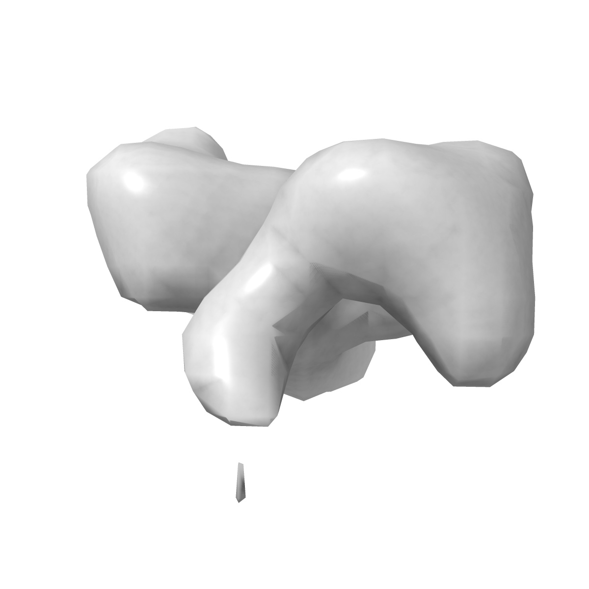
EMD-3708
Negative-stain surface of SorCS1 monomer
EMD-3708
Single-particle18.0 Å
 Deposition: 05/05/2017
Map released: 06/09/2017
Last modified: 01/04/2020
Buffer
pH: 8.0
Staining
Microscope: FEI TECNAI SPIRIT
Illumination mode: FLOOD BEAM
Imaging mode: BRIGHT FIELD
Electron source: LAB6
Acceleration voltage: 120 kV
Illumination mode: FLOOD BEAM
Imaging mode: BRIGHT FIELD
Electron source: LAB6
Acceleration voltage: 120 kV
Image Recording
[1]
Final
reconstruction
Resolution: 18.0
Å
(
BY AUTHOR)
Resolution method: FSC 0.143 CUT-OFF
Number of images used: 34000
Resolution method: FSC 0.143 CUT-OFF
Number of images used: 34000
⌯ Applied Symmetry
Point group:
C1
⦨ Initial angle
assignment
Type:
PROJECTION MATCHING
⦩ Final angle assignment
Type:
PROJECTION MATCHING
Format: CCP4
Data type: IMAGE STORED AS FLOATING POINT NUMBER (4 BYTES)
Annotation details: Negative stain surface of mSorCS1 monomer
Data type: IMAGE STORED AS FLOATING POINT NUMBER (4 BYTES)
Annotation details: Negative stain surface of mSorCS1 monomer
⬡ Geometry
| X | Y | Z | |
|---|---|---|---|
| Dimensions | 28 | 28 | 28 |
| Origin | -14 | -14 | -14 |
| Spacing | 28 | 28 | 28 |
| Voxel size | 6.29 Å | 6.29 Å | 6.29 Å |
Contour list
| Primary | Level | Source |
|---|---|---|
| True | 0.06 | AUTHOR |