EMD-3656
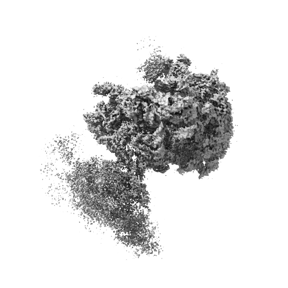
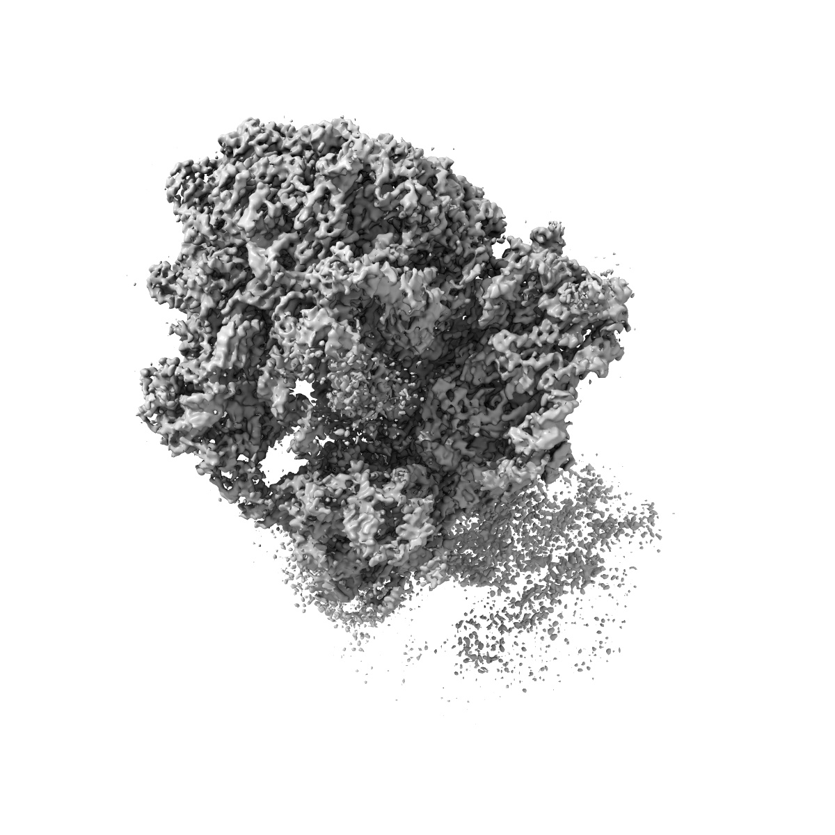
Structure of the Bacillus subtilis hibernating 100S ribosome reveals the basis for 70S dimerization.
EMD-3656
Single-particle3.8 Å
 Deposition: 29/03/2017
Map released: 14/06/2017
Last modified: 20/02/2019
Details: 4 OD260/ml ml Bs100S sample were applied to 2 nm pre-coated Quantifoil R3/3 holey carbon supported grids and vitrified using Vitrobot Mark IV (FEI Company)
Buffer
Grid
Vitrification
Cryogen name: ETHANE
Microscope: FEI TITAN KRIOS
Illumination mode: FLOOD BEAM
Imaging mode: BRIGHT FIELD
Electron source: FIELD EMISSION GUN
Acceleration voltage: 300 kV
Nominal CS: 2.7 mm
Specimen holder model: FEI TITAN KRIOS AUTOGRID HOLDER
Cooling holder cryogen: NITROGEN
Illumination mode: FLOOD BEAM
Imaging mode: BRIGHT FIELD
Electron source: FIELD EMISSION GUN
Acceleration voltage: 300 kV
Nominal CS: 2.7 mm
Specimen holder model: FEI TITAN KRIOS AUTOGRID HOLDER
Cooling holder cryogen: NITROGEN
Image Recording
[1]
Detector model:
FEI FALCON II (4k x 4k)
Detector mode: COUNTING
Average electron dose per image: 2.5 e/Å2
Detector mode: COUNTING
Average electron dose per image: 2.5 e/Å2
Final
reconstruction
Resolution: 3.8
Å
(
BY AUTHOR)
Resolution method: FSC 0.143 CUT-OFF
Number of images used: 24546
Resolution method: FSC 0.143 CUT-OFF
Number of images used: 24546
⌯ Applied Symmetry
Point group:
C1
Software
[1]
| Name | Version | Details |
|---|---|---|
| FREALIGN | 9.11 | - |
⦨ Initial angle
assignment
⦩ Final angle assignment
Particle selection
[1]
| Selected | Ref. model | Method | Software | Details |
|---|---|---|---|---|
| 253905 | - | - | - | - |
Final 3D classification
Software
[1]
| Name | Version | Details |
|---|---|---|
| FREALIGN | 9.11 | - |
CTF correction
Software
[1]
| Name | Version | Details |
|---|---|---|
| CTFFIND | 4 | - |
Format: CCP4
Data type: IMAGE STORED AS FLOATING POINT NUMBER (4 BYTES)
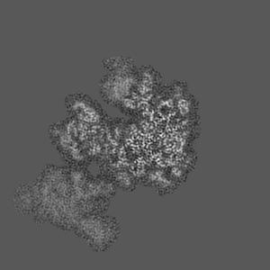
Annotation details: Cryo-EM reconstruction of the Bacillus subtilis 100s subcomplex (70s-30s)
Data type: IMAGE STORED AS FLOATING POINT NUMBER (4 BYTES)
Annotation details: Cryo-EM reconstruction of the Bacillus subtilis 100s subcomplex (70s-30s)
⬡ Geometry
| X | Y | Z | |
|---|---|---|---|
| Dimensions | 450 | 450 | 450 |
| Origin | 0 | 0 | 0 |
| Spacing | 450 | 450 | 450 |
| Voxel size | 1.084 Å | 1.084 Å | 1.084 Å |
Contour list
| Primary | Level | Source |
|---|---|---|
| True | 11.0 | AUTHOR |