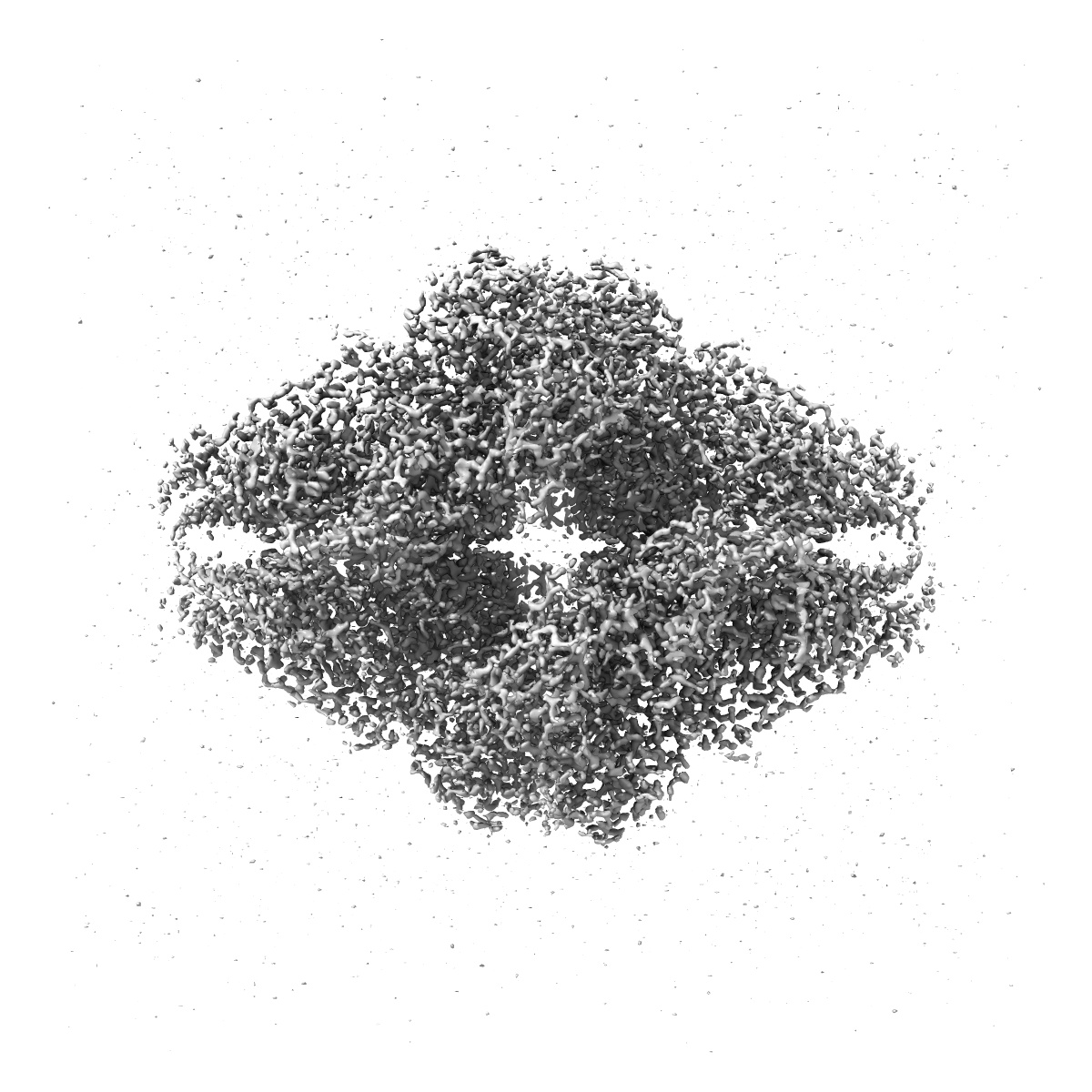
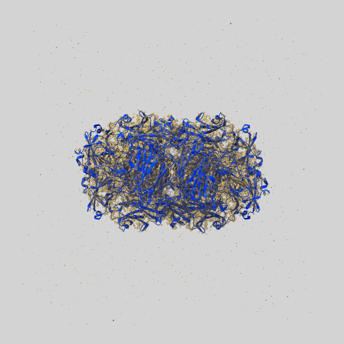
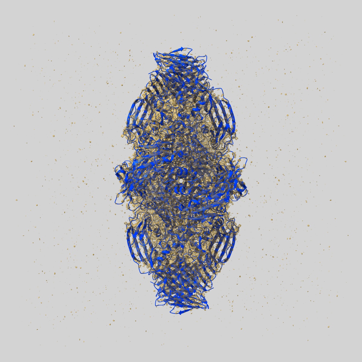
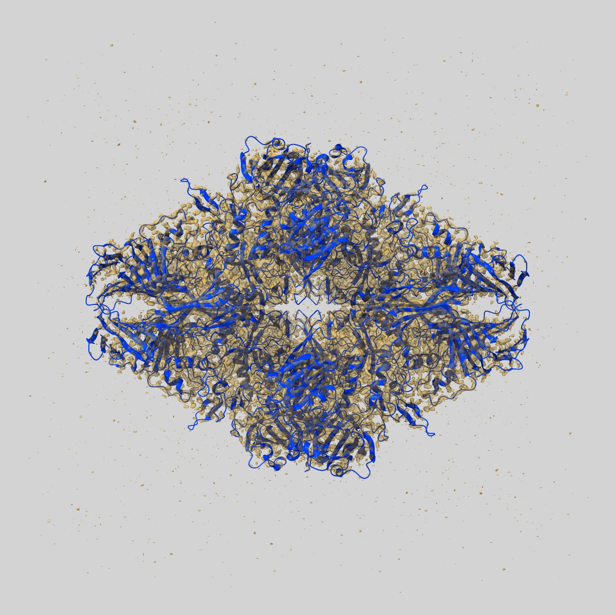
EMD-2984
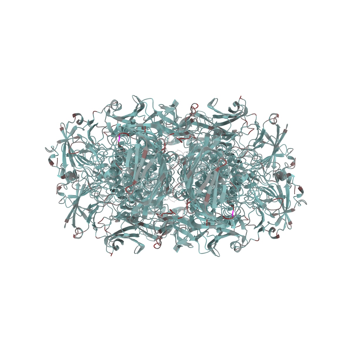
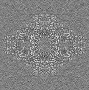
2.2 A resolution cryo-EM structure of beta-galactosidase in complex with a cell-permeant inhibitor
EMD-2984
Single-particle2.2 Å
 Deposition: 26/04/2015
Map released: 06/05/2015
Last modified: 14/10/2015
Name: Escherichia coli beta-galactosidase bound to phenylethyl beta-D-thiogalactopyranoside (PETG)
Summary:
Name: Escherichia coli beta-galactosidase bound to phenylethyl beta-D-thiogalactopyranoside (PETG)
Number of unique components: 2
Oligomeric state: tetramer
Details: The sample was monodisperse.
Number of unique components: 2
Oligomeric state: tetramer
Details: The sample was monodisperse.
Molecular weight
| Experimental | Theoretical | Method |
|---|---|---|
| - | 465 kDa | ProtParam tool: Gasteiger E., Hoogland C., Gattiker A., Duvaud S., Wilkins M.R., Appel R.D., Bairoch A., Protein Identification and Analysis Tools on the ExPASy Server, (in) John M. Walker (ed): The Proteomics Protocols Handbook, Humana Press (2005), pp. 571-607 |
Name: beta-galactosidase
(beta-gal, b-gal)
Number of copies: 1
Oligomeric state: tetramer
UniProtKB PDBe-KB AlphaFold DB
Number of copies: 1
Oligomeric state: tetramer
UniProtKB PDBe-KB AlphaFold DB
Gene Ontology [1]
Natural source
Molecular weight
| Experimental | Theoretical | Method |
|---|---|---|
| - | 465 kDa | - |
Recombinant Expression
| Organism | Strain | Cell | Plasmid |
|---|---|---|---|
| - | - | - | - |
Name: phenylethyl beta-D-thiogalactopyranoside
(PETG)
Number of copies: 4
Oligomeric state: monomer
Details: Molecular weight of PETG is 300.37 Da.
Number of copies: 4
Oligomeric state: monomer
Details: Molecular weight of PETG is 300.37 Da.
Natural source
Organism: unidentified
Recombinant Expression
| Organism | Strain | Cell | Plasmid |
|---|---|---|---|
| - | - | - | - |