EMD-2310
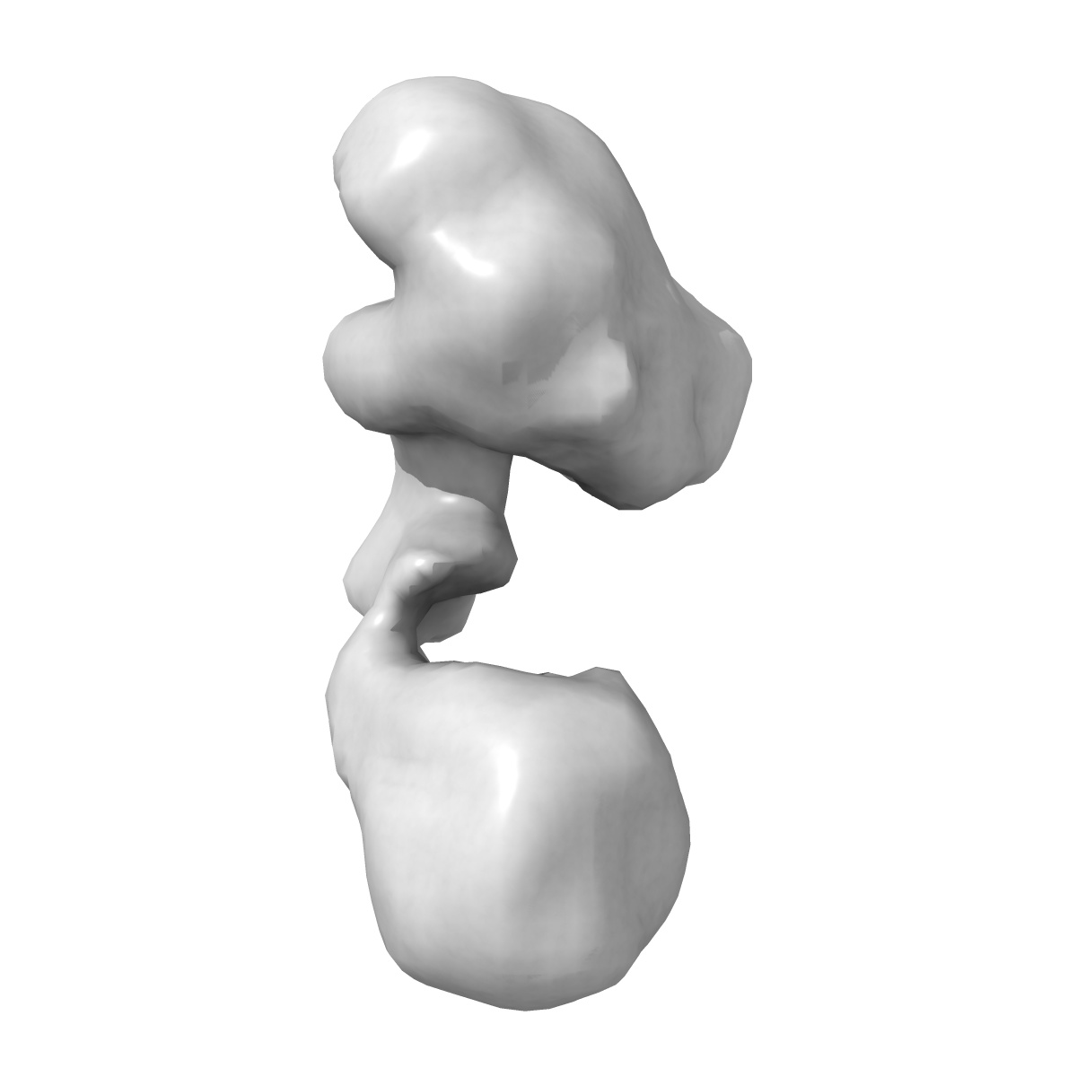
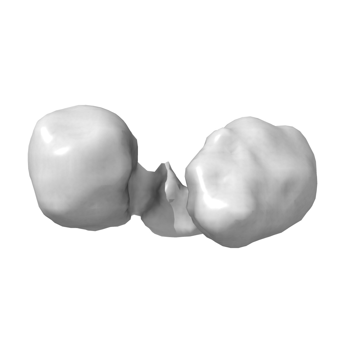
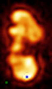
Three-dimensional structure of active, full-length human telomerase dimer, determined by single-particle electron microscopy in negative stain
EMD-2310
Single-particle30.0 Å
 Deposition: 05/02/2013
Map released: 20/03/2013
Last modified: 17/04/2013
Concentration: 0.01
mg/mL
Buffer
pH: 7.6
Details: 20 mM Tris, 150 mM KCl, 1 mM MgCl2
Details: 20 mM Tris, 150 mM KCl, 1 mM MgCl2
Staining
Type:
NEGATIVE
Details: Continuous carbon-coated grids were freshly prepared and glow-discharged before use. 13 microl of telomerase sample (8-10 nM) were deposited on the grid for 15-30 minutes, blotted with filter paper and negatively stained with 2 drops of 1-2% (w/v) uranyl acetate solution.
Details: Continuous carbon-coated grids were freshly prepared and glow-discharged before use. 13 microl of telomerase sample (8-10 nM) were deposited on the grid for 15-30 minutes, blotted with filter paper and negatively stained with 2 drops of 1-2% (w/v) uranyl acetate solution.
Grid
Details: 200 mesh carbon coated with thin carbon, glow discharged
Microscope: FEI TECNAI 12
Illumination mode: FLOOD BEAM
Imaging mode: BRIGHT FIELD
Electron source: TUNGSTEN HAIRPIN
Acceleration voltage: 120 kV
Nominal defocus: 0.001 µm - 0.0015 µm
Nominal magnification: 42000.0
Calibrated magnification: 42550.0
Specimen holder model: SIDE ENTRY, EUCENTRIC
Alignment procedure: LEGACY (Astigmatism: Corrected at 100,000 times magnification, Electron beam tilt params: )
Details: The films were developed in Kodak developer at full strength for 12 min.
Image Recording
[1]
Detector category:
FILM
Detector model: KODAK SO-163 FILM
Scanner: ZEISS SCAI
Sampling interval: 7 µm
Number of real images: 482
Average electron dose per image: 10 e/Å2
Old range: 1.4
Details: The micrographs were compressed x4
Detector model: KODAK SO-163 FILM
Scanner: ZEISS SCAI
Sampling interval: 7 µm
Number of real images: 482
Average electron dose per image: 10 e/Å2
Old range: 1.4
Details: The micrographs were compressed x4
Details: Electron tomography was used to calculate a low-resolution reference model. Distinct conformations were further classified by three-dimensional maximum-likelihood analysis in Fourier space (MLF3D).Density map calculated from a sub-population of 3,659 particles.
Final
reconstruction
Resolution: 30.0
Å
(
BY AUTHOR)
Resolution method: OTHER
Number of images used: 20127
Algorithm: OTHER
Details: 3D structure was refined using the low-resolution subtomogram average as a reference model for 3D single particle refinement with EMAN2. Distinct conformations were further classified by three-dimensional maximum-likelihood analysis in Fourier space (MLF3D). Density map calculated from a sub-population of 3,659 particles. The absolute hand and the overall correctness of the dimer structure were assessed using a modified version of tilt-pair validation method.
⌯ Applied Symmetry
Point group:
C1
Software
[1]
| Name | Version | Details |
|---|---|---|
| EMAN2, XMIPP | - | - |
Final 2D classification
Number of classes:
100
Format: CCP4
Data type: IMAGE STORED AS FLOATING POINT NUMBER (4 BYTES)
Annotation details: 3D map of human telomerase dimer bound to G-overhang oligonucleotide (TTAGGG)2
Details: ::::EMDATABANK.org::::EMD-2310::::
Data type: IMAGE STORED AS FLOATING POINT NUMBER (4 BYTES)
Annotation details: 3D map of human telomerase dimer bound to G-overhang oligonucleotide (TTAGGG)2
Details: ::::EMDATABANK.org::::EMD-2310::::
⬡ Geometry
| X | Y | Z | |
|---|---|---|---|
| Dimensions | 48 | 28 | 28 |
| Origin | 17 | 26 | 25 |
| Spacing | 28 | 48 | 28 |
| Voxel size | 6.6 Å | 6.6 Å | 6.6 Å |
Contour list
| Primary | Level | Source |
|---|---|---|
| True | 0.0974 | AUTHOR |