EMD-1648
Assembly and Allosteric Mechanism of Molluscan Hemocyanin Revealed by Cryo-EM Structure and Pseudo-atomic Model
EMD-1648
Single-particle7.8 Å
 Deposition: 10/09/2009
Map released: 12/11/2010
Last modified: 25/05/2016
Buffer
pH: 7.5
Details: 0.2M NaCl , 50mM Tris-HCl, 5mM CaCl2, 5mM MgCl2, pH7.5
Details: 0.2M NaCl , 50mM Tris-HCl, 5mM CaCl2, 5mM MgCl2, pH7.5
Grid
Details: 1.2/1.3 copper Quantifoil grids
Microscope: JEOL 3200FSC
Illumination mode: FLOOD BEAM
Imaging mode: BRIGHT FIELD
Electron source: FIELD EMISSION GUN
Acceleration voltage: 300 kV
Nominal CS: 4.1 mm
Nominal defocus: 0.8 µm - 3.5 µm
Nominal magnification: 60000.0
Specimen holder model: JEOL 3200FSC CRYOHOLDER
Specimen holder details: Eucentric
Alignment procedure: LEGACY (Astigmatism: Objective lens astigmatism was corrected at 400,000 times magnification, Electron beam tilt params: )
Details: MDS
Temperature
Average: 101
K
Specialist optics
Energy filter
Name:
JEOL in-column
Image Recording
[1]
Detector category:
FILM
Detector model: KODAK SO-163 FILM
Scanner: NIKON SUPER COOLSCAN 9000
Sampling interval: 6.35 µm
Number of real images: 820
Average electron dose per image: 18 e/Å2
Bits per pixel: 16.0
Detector model: KODAK SO-163 FILM
Scanner: NIKON SUPER COOLSCAN 9000
Sampling interval: 6.35 µm
Number of real images: 820
Average electron dose per image: 18 e/Å2
Bits per pixel: 16.0
Details: Particles were automatically boxed out from micrographs by e2boxer.py from EMAN2 single particle analysis software package, and CTF correction was carried out with EMAN program CTFIT. All the 3D reconstruction was done with EMAN1.8.
Final
reconstruction
Resolution: 7.8
Å
(
BY AUTHOR)
Resolution method: FSC 0.5 CUT-OFF
Number of images used: 41650
Details:
Resolution method: FSC 0.5 CUT-OFF
Number of images used: 41650
Details:
⌯ Applied Symmetry
Point group:
D5
Software
[1]
| Name | Version | Details |
|---|---|---|
| EMAN | - | - |
CTF correction
Details:Each micrograph
Format: CCP4
Data type: IMAGE STORED AS FLOATING POINT NUMBER (4 BYTES)
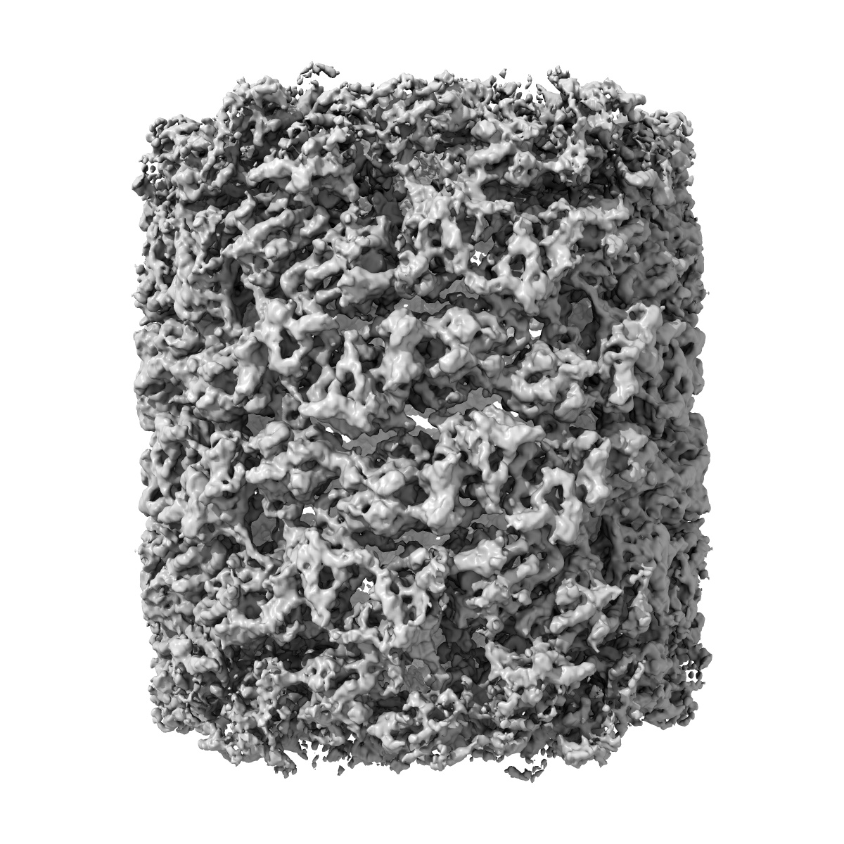
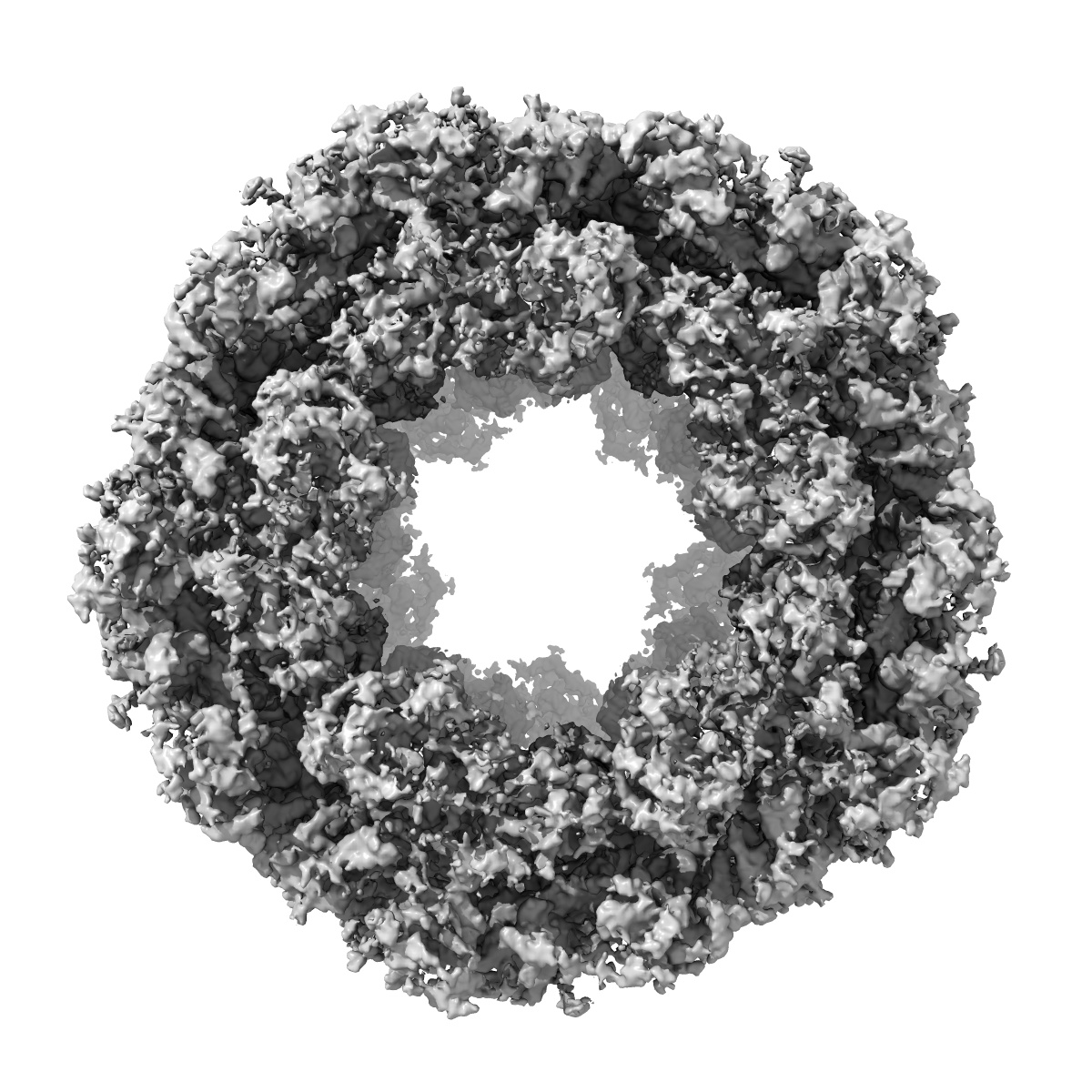
Annotation details: The whole structure of Haliotis diversicolor Hemocyanin isoform 1 (HdH1) is a hollow cylindrical dodecamer. Each of its 20 subunits is composed of 8 functional units (FUs).
Details: ::::EMDATABANK.org::::EMD-1648::::
Data type: IMAGE STORED AS FLOATING POINT NUMBER (4 BYTES)
Annotation details: The whole structure of Haliotis diversicolor Hemocyanin isoform 1 (HdH1) is a hollow cylindrical dodecamer. Each of its 20 subunits is composed of 8 functional units (FUs).
Details: ::::EMDATABANK.org::::EMD-1648::::
⬡ Geometry
| X | Y | Z | |
|---|---|---|---|
| Dimensions | 432 | 432 | 432 |
| Origin | -216 | -216 | -216 |
| Spacing | 432 | 432 | 432 |
| Voxel size | 1.06 Å | 1.06 Å | 1.06 Å |
Contour list
| Primary | Level | Source |
|---|---|---|
| True | 1.37 | EMDB |