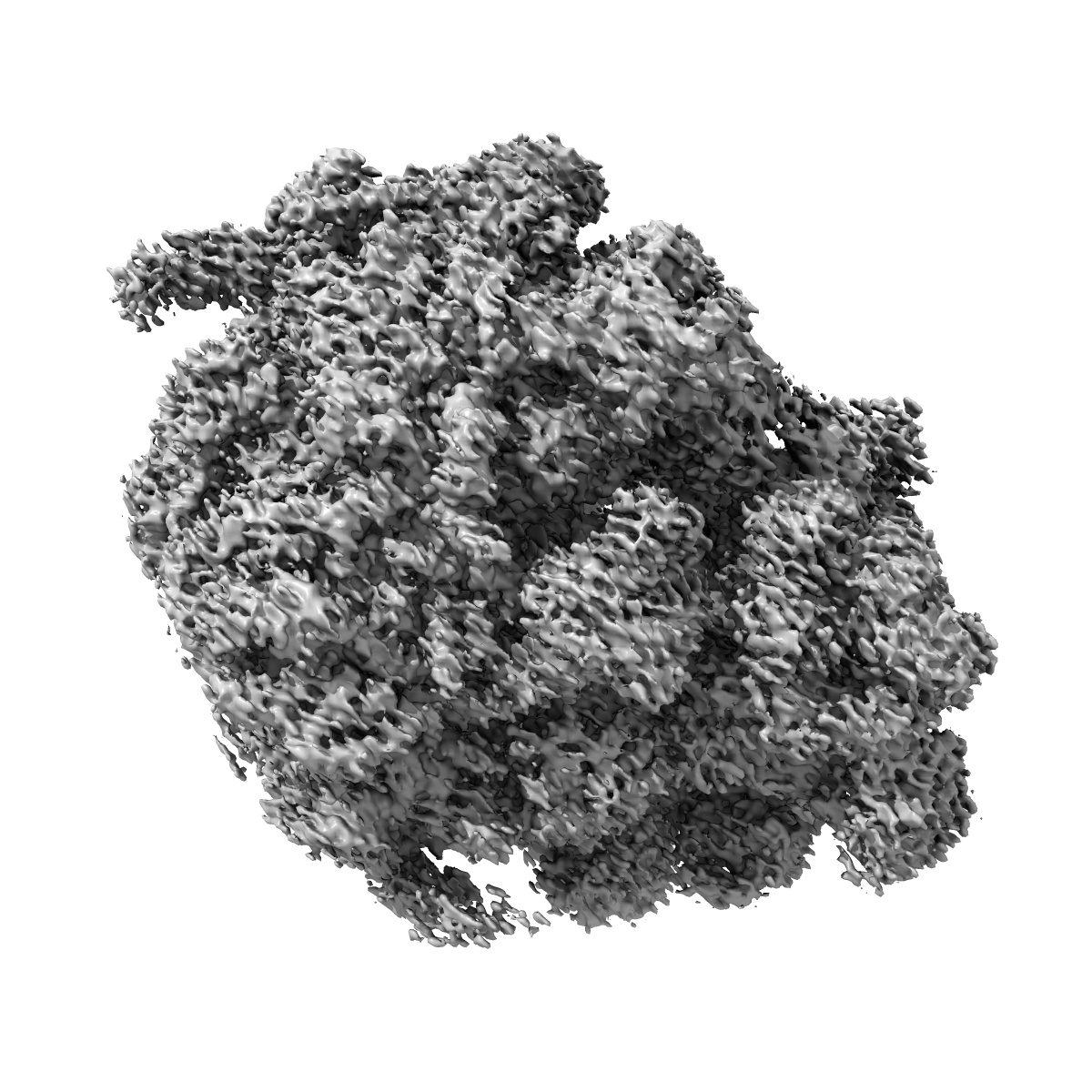
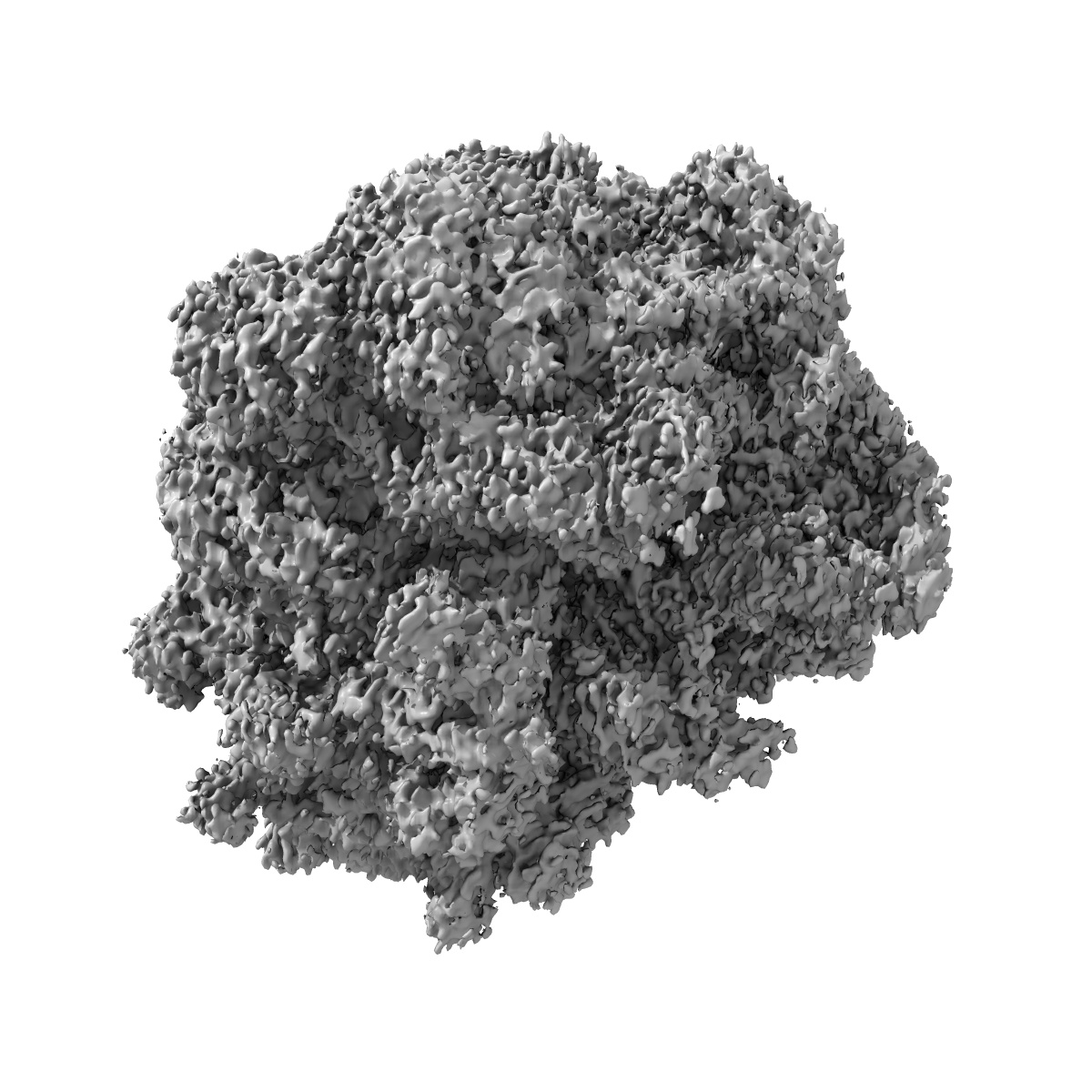
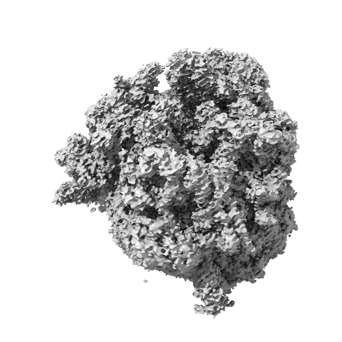
EMD-0049
Cryo-EM recosntruction of yeast 80S ribosome in complex with mRNA, tRNA and eEF2 (GMPPCP)
EMD-0049
Single-particle4.0 Å
 Deposition: 08/06/2018
Map released: 11/07/2018
Last modified: 08/08/2018
Buffer
Vitrification
Cryogen name: ETHANE
Chamber humidity: 100%
Chamber temperature: 277 K
Instrument: FEI VITROBOT MARK IV
Details: Blot force 4, blot waiting time 30 s.
Chamber humidity: 100%
Chamber temperature: 277 K
Instrument: FEI VITROBOT MARK IV
Details: Blot force 4, blot waiting time 30 s.
Microscope: FEI TITAN KRIOS
Illumination mode: FLOOD BEAM
Imaging mode: BRIGHT FIELD
Electron source: FIELD EMISSION GUN
Acceleration voltage: 300 kV
Illumination mode: FLOOD BEAM
Imaging mode: BRIGHT FIELD
Electron source: FIELD EMISSION GUN
Acceleration voltage: 300 kV
Image Recording
[1]
Detector model:
FEI FALCON II (4k x 4k)
Average exposure time: 1.5 s
Average electron dose per image: 60.0 e/Å2
Average exposure time: 1.5 s
Average electron dose per image: 60.0 e/Å2
Final
reconstruction
Resolution: 4.0
Å
(
BY AUTHOR)
Resolution method: FSC 0.143 CUT-OFF
Number of images used: 86500
Resolution method: FSC 0.143 CUT-OFF
Number of images used: 86500
⌯ Applied Symmetry
Point group:
C1
Software
[1]
| Name | Version | Details |
|---|---|---|
| RELION | - | - |
⦨ Initial angle
assignment
Type:
MAXIMUM LIKELIHOOD
⦩ Final angle assignment
Type:
MAXIMUM LIKELIHOOD
Format: CCP4
Data type: IMAGE STORED AS FLOATING POINT NUMBER (4 BYTES)
Annotation details: None
Data type: IMAGE STORED AS FLOATING POINT NUMBER (4 BYTES)
Annotation details: None
⬡ Geometry
| X | Y | Z | |
|---|---|---|---|
| Dimensions | 360 | 360 | 360 |
| Origin | 0 | 0 | 0 |
| Spacing | 360 | 360 | 360 |
| Voxel size | 1.1 Å | 1.1 Å | 1.1 Å |
Contour list
| Primary | Level | Source |
|---|---|---|
| True | 0.033 | EMDB |