EMD-2479
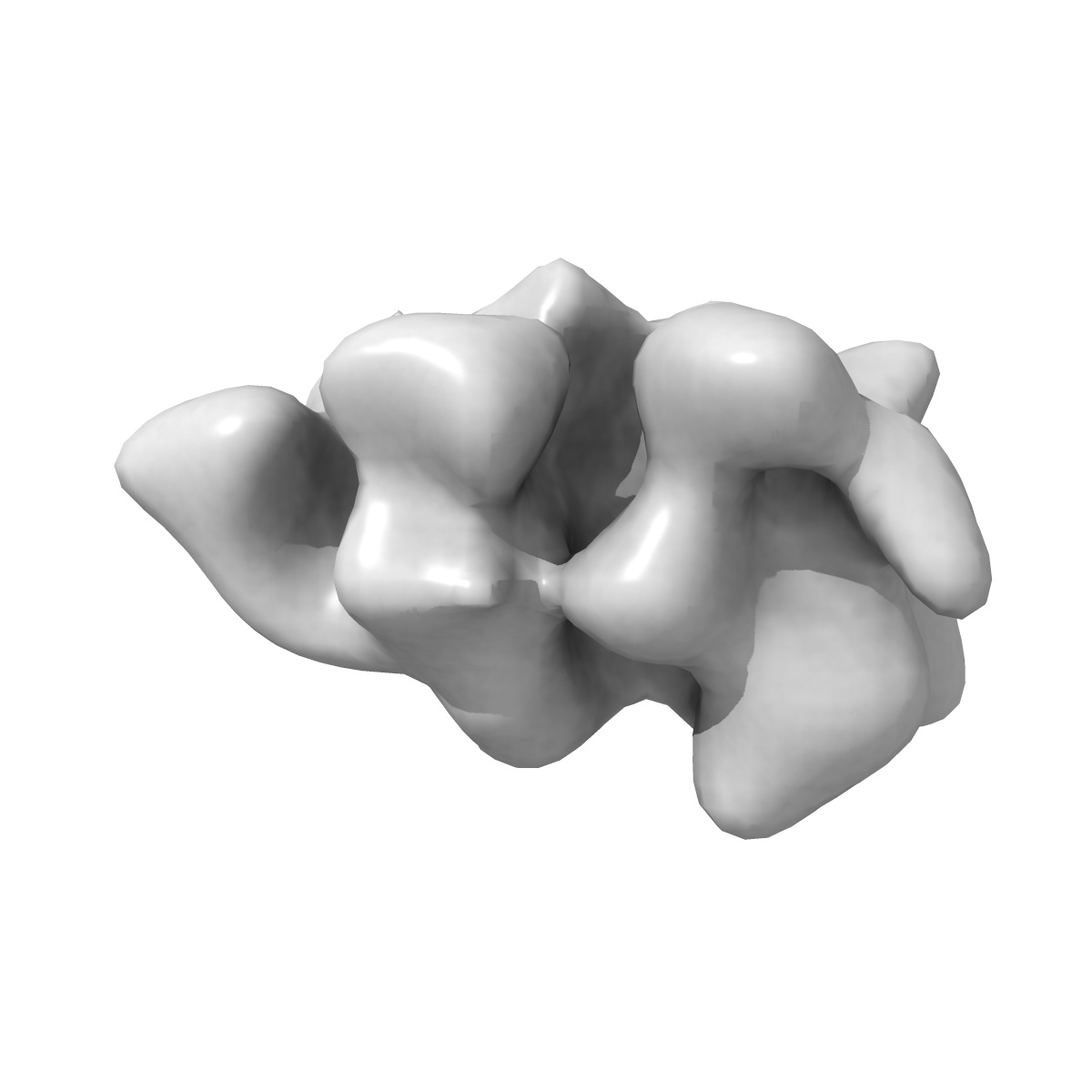
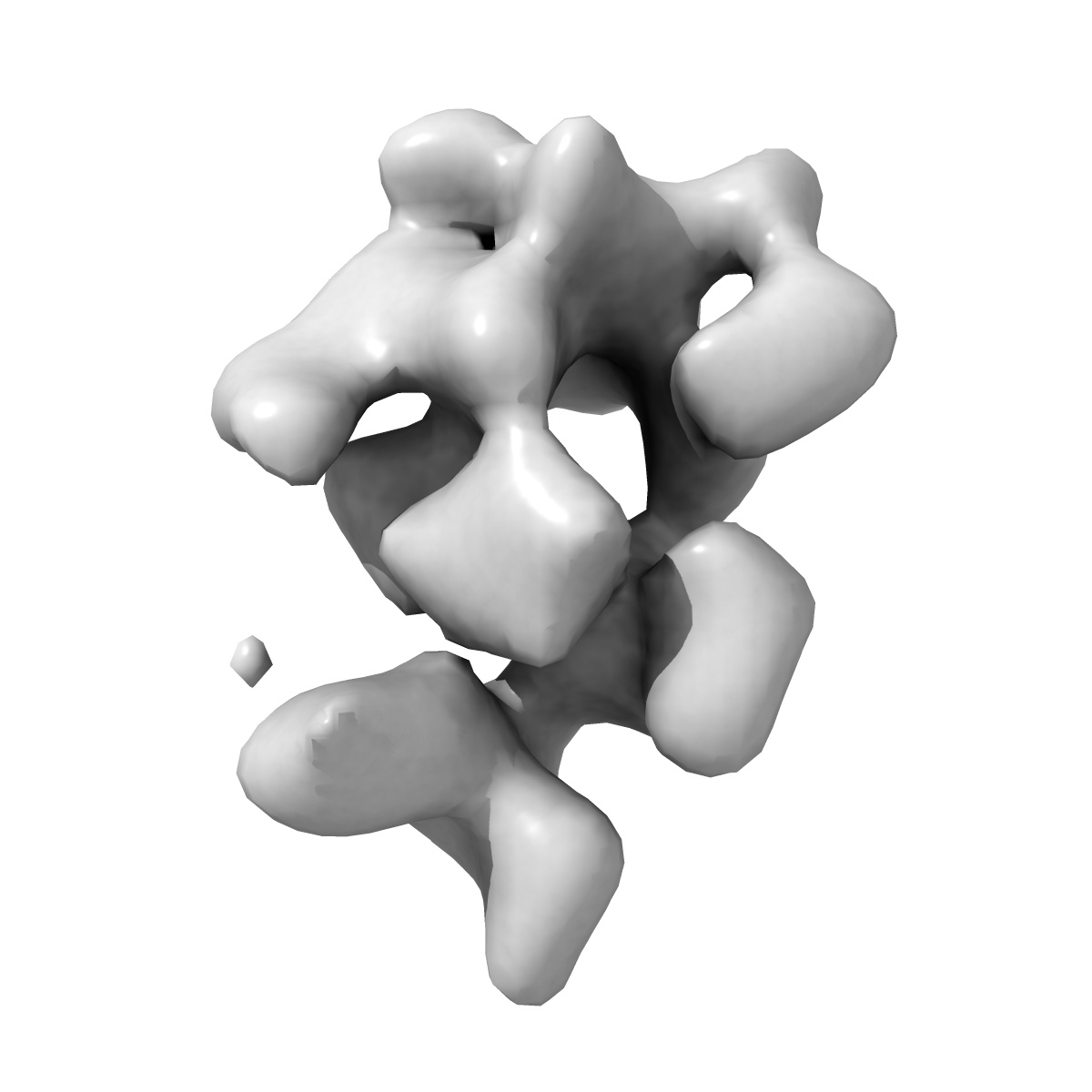
Electron microscopy structure of the Drosophila origin recognition complex
EMD-2479
Single-particle22.0 Å
 Deposition: 26/09/2013
Map released: 30/10/2013
Last modified: 30/10/2013
Concentration: 0.0116
mg/mL
Buffer
pH: 7.8
Details: 50mM Tris-HCL pH 7.8, 300mM KCl, 5mM MgCl2, 1mM ATPgS
Details: 50mM Tris-HCL pH 7.8, 300mM KCl, 5mM MgCl2, 1mM ATPgS
Staining
Type:
NEGATIVE
Details: Grids with adsorbed protein were floated on 4 drops of 2% uranyl formate for 10 seconds each
Details: Grids with adsorbed protein were floated on 4 drops of 2% uranyl formate for 10 seconds each
Grid
Details: 400 mesh copper grid with continuous carbon support
Microscope: FEI TECNAI 12
Illumination mode: FLOOD BEAM
Imaging mode: BRIGHT FIELD
Electron source: LAB6
Acceleration voltage: 120 kV
Nominal CS: 6.3 mm
Nominal defocus: 0.4 µm - 1.2 µm
Nominal magnification: 49000.0
Specimen holder model: SIDE ENTRY, EUCENTRIC
Minimum tilt angle: 0
Maximum tilt angle: 30
Illumination mode: FLOOD BEAM
Imaging mode: BRIGHT FIELD
Electron source: LAB6
Acceleration voltage: 120 kV
Nominal CS: 6.3 mm
Nominal defocus: 0.4 µm - 1.2 µm
Nominal magnification: 49000.0
Specimen holder model: SIDE ENTRY, EUCENTRIC
Minimum tilt angle: 0
Maximum tilt angle: 30
Temperature
Average: 297
K
Image Recording
[1]
Detector category:
CCD
Detector model: TVIPS TEMCAM-F416 (4k x 4k)
Average electron dose per image: 25 e/Å2
Detector model: TVIPS TEMCAM-F416 (4k x 4k)
Average electron dose per image: 25 e/Å2
Details: Particles were selected using the automated particle selection software DoG Picker. 3D reconstructions were performed with SPIDER.
Final
reconstruction
Resolution: 22.0
Å
(
BY AUTHOR)
Resolution method: FSC 0.5 CUT-OFF
Number of images used: 70000
Algorithm: OTHER
Details: Particles were automatically selected with DoG Picker. The contrast transfer function was estimated with CTFFIND/CTFTILT and phases flipped with SPIDER. Projection-matching refinement was performed with SPIDER using a previously determined 3D reconstruction of Drosophila ORC as a starting model.
⌯ Applied Symmetry
Point group:
C1
Software
[1]
| Name | Version | Details |
|---|---|---|
| SPIDER | - | - |
CTF correction
Details:Each micrograph for untilted, each particle for tilted
Format: CCP4
Data type: IMAGE STORED AS FLOATING POINT NUMBER (4 BYTES)
Annotation details: Reconstruction of Drosophila ORC in presence of 1mM ATPgS
Details: ::::EMDATABANK.org::::EMD-2479::::
Data type: IMAGE STORED AS FLOATING POINT NUMBER (4 BYTES)
Annotation details: Reconstruction of Drosophila ORC in presence of 1mM ATPgS
Details: ::::EMDATABANK.org::::EMD-2479::::
⬡ Geometry
| X | Y | Z | |
|---|---|---|---|
| Dimensions | 80 | 80 | 80 |
| Origin | -40 | -40 | -40 |
| Spacing | 80 | 80 | 80 |
| Voxel size | 4.36 Å | 4.36 Å | 4.36 Å |
Contour list
| Primary | Level | Source |
|---|---|---|
| True | 4.5 | AUTHOR |