EMD-2321
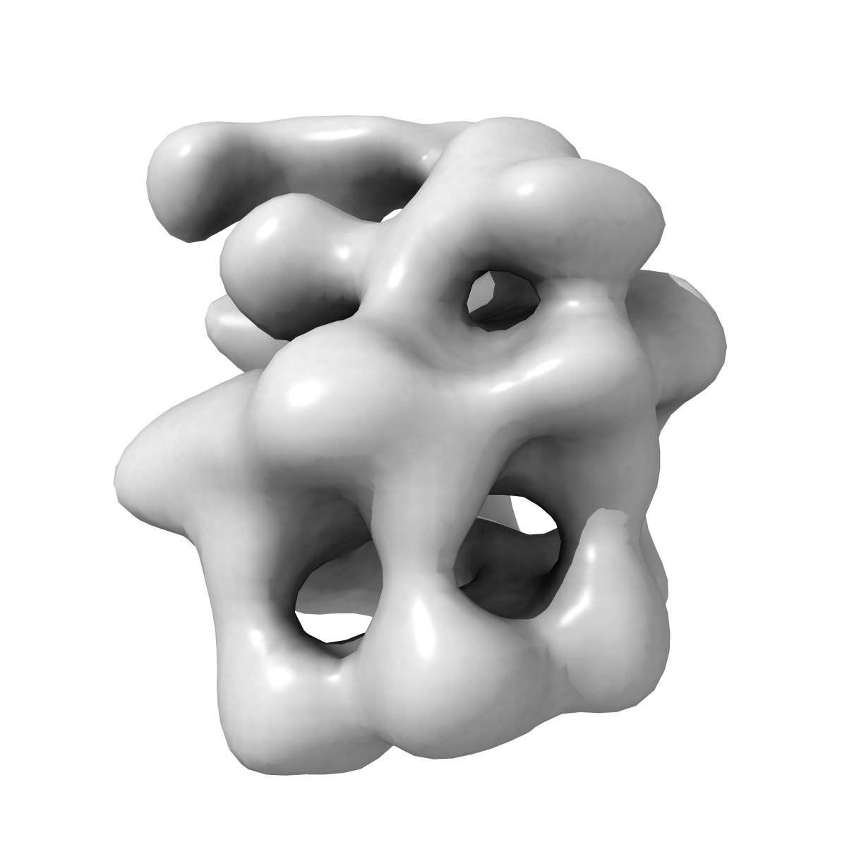
The bacterial DnaC helicase loader is a DnaB ring breaker
EMD-2321
Single-particle25.0 Å
 Deposition: 26/02/2013
Map released: 17/04/2013
Last modified: 24/04/2013
Buffer
pH: 8.5
Details: 20 mM Tris-HCl pH 8.5, 200 mM NaCl, 5 % glycerol, 5 mM MgCl2, 1 mM beta-mercaptoethanol, and 1 mM ADP-BeF3
Details: 20 mM Tris-HCl pH 8.5, 200 mM NaCl, 5 % glycerol, 5 mM MgCl2, 1 mM beta-mercaptoethanol, and 1 mM ADP-BeF3
Staining
Type:
NEGATIVE
Details: Grids with adsorbed protein were floated on 2% w/v uranyl formate for 45 seconds
Details: Grids with adsorbed protein were floated on 2% w/v uranyl formate for 45 seconds
Grid
Details: 400 mesh copper grid with thin carbon support, glow discharged for 20 seconds
Microscope: FEI TECNAI 12
Illumination mode: FLOOD BEAM
Imaging mode: BRIGHT FIELD
Electron source: LAB6
Acceleration voltage: 120 kV
Nominal CS: 6.3 mm
Nominal defocus: 0.7 µm - 1.2 µm
Nominal magnification: 49000.0
Specimen holder model: SIDE ENTRY, EUCENTRIC
Illumination mode: FLOOD BEAM
Imaging mode: BRIGHT FIELD
Electron source: LAB6
Acceleration voltage: 120 kV
Nominal CS: 6.3 mm
Nominal defocus: 0.7 µm - 1.2 µm
Nominal magnification: 49000.0
Specimen holder model: SIDE ENTRY, EUCENTRIC
Temperature
Average: 297
K
Image Recording
[1]
Detector category:
CCD
Detector model: GENERIC TVIPS (4k x 4k)
Average electron dose per image: 25 e/Å2
Detector model: GENERIC TVIPS (4k x 4k)
Average electron dose per image: 25 e/Å2
Details: The particles were selected using DoG picker as available in APPION. The contrast transfer function of the microscope for each micrograph was estimated using CTFFIND3 and phase-flipped using SPIDER. DnaBC particles were subjected to a multi-model refinement as implemented in SPARX using the 3D averages obtained from the RCT reconstructions as initial references.
Final
reconstruction
Resolution: 25.0
Å
(
BY AUTHOR)
Resolution method: FSC 0.5 CUT-OFF
Number of images used: 17942
Algorithm: OTHER
Details:
Resolution method: FSC 0.5 CUT-OFF
Number of images used: 17942
Algorithm: OTHER
Details:
⌯ Applied Symmetry
Point group:
C1
Software
[1]
| Name | Version | Details |
|---|---|---|
| EMAN2, SPARX | - | - |
CTF correction
Details:Each micrograph
Format: CCP4
Data type: IMAGE STORED AS FLOATING POINT NUMBER (4 BYTES)
Annotation details: Negative staining reconstruction of E. coli DnaB/DnaC complex
Details: ::::EMDATABANK.org::::EMD-2321::::
Data type: IMAGE STORED AS FLOATING POINT NUMBER (4 BYTES)
Annotation details: Negative staining reconstruction of E. coli DnaB/DnaC complex
Details: ::::EMDATABANK.org::::EMD-2321::::
⬡ Geometry
| X | Y | Z | |
|---|---|---|---|
| Dimensions | 80 | 80 | 80 |
| Origin | 0 | 0 | 0 |
| Spacing | 80 | 80 | 80 |
| Voxel size | 4.36 Å | 4.36 Å | 4.36 Å |
Contour list
| Primary | Level | Source |
|---|---|---|
| True | 3.0 | AUTHOR |