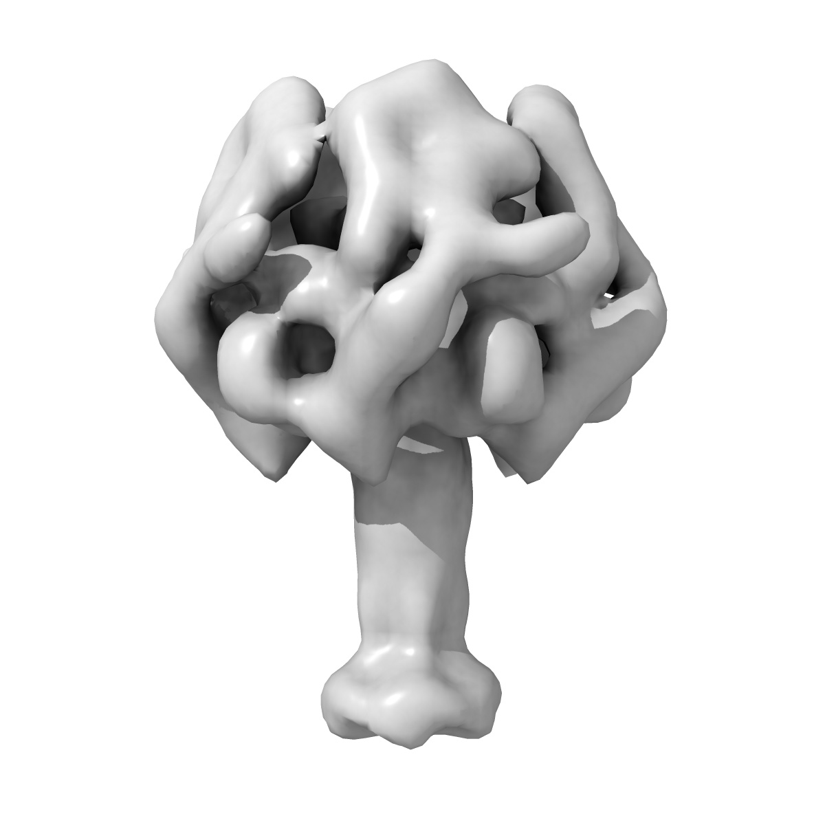
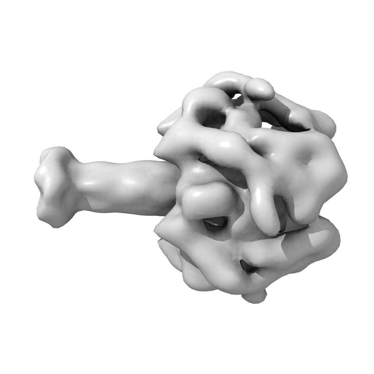
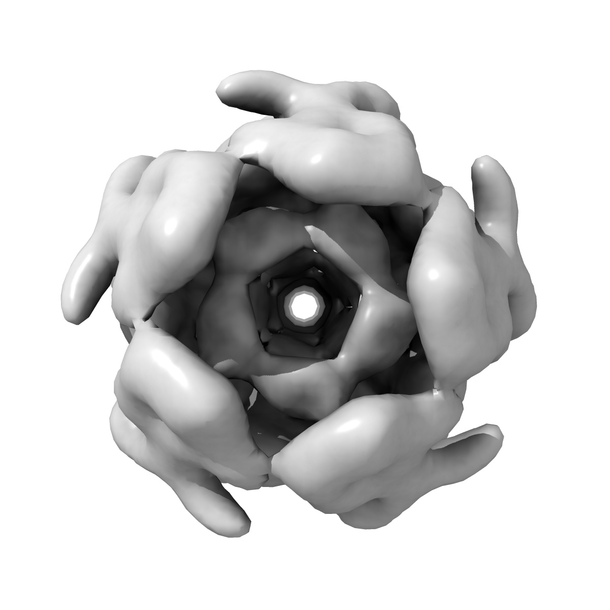
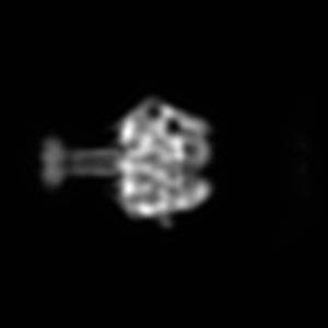
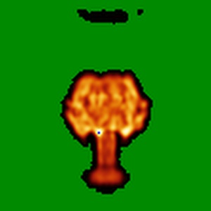
EMD-2301
Negative stain EM structure of TcdA1 in pore state
EMD-2301
Single-particle26.5 Å
 Deposition: 25/01/2013
Map released: 13/02/2013
Last modified: 03/04/2013
Concentration: 0.02
mg/mL
Buffer
pH: 11.0
Details: 50 mM CAPS, mM NaCl, 0.05% Tween-20, 5% glycerol
Details: 50 mM CAPS, mM NaCl, 0.05% Tween-20, 5% glycerol
Staining
Type:
NEGATIVE
Details: Grids with adsorbed Protein floated on 0.07% Uranyl Formate for 45 sec.
Details: Grids with adsorbed Protein floated on 0.07% Uranyl Formate for 45 sec.
Grid
Details: Glow discharged 400 mesh copper grids with thin carbon support
Microscope: JEOL 1400
Illumination mode: FLOOD BEAM
Imaging mode: BRIGHT FIELD
Electron source: LAB6
Acceleration voltage: 120 kV
Nominal CS: 3.4 mm
Nominal defocus: 0.0012 µm - 0.002 µm
Nominal magnification: 50000.0
Calibrated magnification: 67210.0
Specimen holder model: JEOL
Illumination mode: FLOOD BEAM
Imaging mode: BRIGHT FIELD
Electron source: LAB6
Acceleration voltage: 120 kV
Nominal CS: 3.4 mm
Nominal defocus: 0.0012 µm - 0.002 µm
Nominal magnification: 50000.0
Calibrated magnification: 67210.0
Specimen holder model: JEOL
Image Recording
[1]
Final
reconstruction
Resolution: 26.5
Å
(
BY AUTHOR)
Resolution method: FSC 0.5 CUT-OFF
Number of images used: 1936
Algorithm: OTHER
Resolution method: FSC 0.5 CUT-OFF
Number of images used: 1936
Algorithm: OTHER
⌯ Applied Symmetry
Point group:
C5
Software
[1]
| Name | Version | Details |
|---|---|---|
| Sparx | - | - |
Format: CCP4
Data type: IMAGE STORED AS FLOATING POINT NUMBER (4 BYTES)
Annotation details: Negative stain reconstruction of TcdA1 in pore state
Details: ::::EMDATABANK.org::::EMD-2301::::
Data type: IMAGE STORED AS FLOATING POINT NUMBER (4 BYTES)
Annotation details: Negative stain reconstruction of TcdA1 in pore state
Details: ::::EMDATABANK.org::::EMD-2301::::
⬡ Geometry
| X | Y | Z | |
|---|---|---|---|
| Dimensions | 112 | 112 | 112 |
| Origin | 0 | 0 | 0 |
| Spacing | 112 | 112 | 112 |
| Voxel size | 4.64 Å | 4.64 Å | 4.64 Å |
Contour list
| Primary | Level | Source |
|---|---|---|
| True | 0.038 | AUTHOR |