It is estimated that around 37 million people are living with an HIV infection. The molecule on the surface of HIV that recognises host cells and enables the virus to get inside them is depicted in our calendar image for January.
The env gene of HIV encodes a protein called gp160. Gp160 associates with two other copies of itself to form trimers on the surface of the viral membrane, a location in which it can interact with the outside world. Gp160 is cleaved as the virus is produced to form two mature proteins, gp120 and gp41. These remain together and enable to virus to find and stick to the cells it infects, and to get inside them. This is the first stage in the virus replicating itself- because HIV, like all viruses, needs to get inside a cell and ‘hijack’ its machinery to reproduce. Gp120 is responsible for binding to a specific receptor on human cell surfaces (a protein called CD4). Gp41 on the other hand drives the huge changes in shape needed to fuse the viral and host membranes. Ebola glycoprotein, a previous featured structure, serves the same function to recognise host cells and internalise the virus.
At the centre of the glyoprotein trimer is a helix which is part of gp41. It is around this helix that the trimer is organised and which serves as an anchor around which the protein alters its shape- like a transformer toy- on fusing with the host cell membrane.
Because the envelope glycoprotein is critical for infection, it is an obvious target for HIV therapy and thus the subject of much research. If the body can produce antibodies to the envelope glycoprotein, the virus can be prevented from binding to, and infecting human cells. Vaccines could aid in raising antibodies, but unfortunately, the envelope glycoprotein is extremely variable due to the high mutation rate of the virus, and also masked by extensive glycosylation, both features which make raising antibodies difficult. There are many structures in the PDB of fragments of glycoprotein bound to antibodies, in efforts to understand the their interaction. While there are many promising studies on creating an HIV vaccine, some in clinical trials, no vaccine is yet available. The HIV envelope glycoprotein is a challenging protein to study by X-ray crystallography as it sits in a membrane, it is heavily glycosylated and conformationally heterogenous. Electron microscopy is better placed to study proteins with these characteristics and the image in our calendar is based on the cryoEM map of the envelope glycoprotein solved by Sriram Subramaniam's group. The ap is available in our sister archive EMDB. with code EMD-2482 and the atomic model in the PDB is code 4cc8
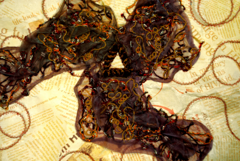
The art was created by Anna Valchanova, a former student at Stephen Perse School in Cambridge, UK. The protein is depicted in layers of yarn. Each layer is abstracted further from the original structure underneath the map of organza fabric, reflecting the high mutation rate that is typical of HIV. This is set against a backdrop of newspaper clippings documenting the social impacts of this virus. At the centre of the envelope glycoprotein are the three important and conserved helices.