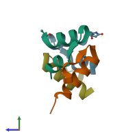
6rlx

X-ray diffraction
1.5Å resolution

X-RAY STRUCTURE OF HUMAN RELAXIN AT 1.5 ANGSTROMS. COMPARISON TO INSULIN AND IMPLICATIONS FOR RECEPTOR BINDING DETERMINANTS

Released:

Function and Biology Details

Biochemical function:
  • not assigned
Biological process:
  • not assigned
Cellular component:
  • not assigned

Structure analysis Details

Assembly composition:
hetero tetramer (preferred)
Assembly name:
PDBe Complex ID:
PDB-CPX-137402 (preferred)
Entry contents:
2 distinct polypeptide molecules
Macromolecules (2 distinct):
Relaxin A chain Chains: A, C
Molecule details ›
Chains: A, C
Length: 24 amino acids
Theoretical weight: 2.66 KDa
Source organism: Homo sapiens
Expression system: Escherichia coli
UniProt:
  • Canonical: P04090 (Residues: 162-185; Coverage: 15%)
Gene name: RLN2
Relaxin B chain Chains: B, D
Molecule details ›
Chains: B, D
Length: 28 amino acids
Theoretical weight: 3.2 KDa
Source organism: Homo sapiens
Expression system: Escherichia coli
UniProt:
  • Canonical: P04090 (Residues: 26-53; Coverage: 17%)
Gene name: RLN2

Ligands and Environments

No bound ligands
1 modified residue:

Experiments and Validation Details

Entry percentile scores
Spacegroup: C2221
Unit cell:
a: 53.97Å b: 56.88Å c: 61.68Å
α: 90° β: 90° γ: 90°
R-values:
R R work R free
0.187 0.187 not available
Expression system: Escherichia coli