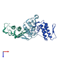
4h0s

X-ray diffraction
1.55Å resolution

Crystal structure analysis of a basic phospholipase A2 from Trimeresurus stejnegeri venom

Released:
Source organism: Trimeresurus stejnegeri
Entry authors: Zeng F, Niu L, Li X, Teng M

Function and Biology Details

Reaction catalysed:
Phosphatidylcholine + H(2)O = 1-acylglycerophosphocholine + a carboxylate
Biochemical function:
Biological process:
Cellular component:
  • not assigned

Structure analysis Details

Assembly composition:
monomeric (preferred)
PDBe Complex ID:
PDB-CPX-179728 (preferred)
Entry contents:
1 distinct polypeptide molecule
Macromolecule:
Basic phospholipase A2 homolog CTs-R6 Chains: A, B, C
Molecule details ›
Chains: A, B, C
Length: 137 amino acids
Theoretical weight: 15.32 KDa
Source organism: Trimeresurus stejnegeri
UniProt:
  • Canonical: Q6H3D7 (Residues: 1-137; Coverage: 100%)
Sequence domains: Phospholipase A2
Structure domains: Phospholipase A2 domain

Ligands and Environments

1 bound ligand:
No modified residues

Experiments and Validation Details

Entry percentile scores
X-ray source: RIGAKU RUH3R
Spacegroup: C2
Unit cell:
a: 95.946Å b: 71.436Å c: 53.607Å
α: 90° β: 103.53° γ: 90°
R-values:
R R work R free
0.183 0.181 0.223