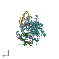
4l5n

X-ray diffraction
2.16Å resolution

Crystallographic Structure of HHV-1 Uracil-DNA Glycosylase complexed with the Bacillus phage PZA inhibitor protein p56

Released:

Function and Biology Details

Reaction catalysed:
Hydrolyzes single-stranded DNA or mismatched double-stranded DNA and polynucleotides, releasing free uracil
Biological process:
Cellular component:
  • not assigned

Structure analysis Details

Assemblies composition:
hetero trimer (preferred)
monomeric
homo dimer
PDBe Complex ID:
PDB-CPX-139309 (preferred)
Entry contents:
2 distinct polypeptide molecules
Macromolecules (2 distinct):
Uracil-DNA glycosylase Chains: A, B
Molecule details ›
Chains: A, B
Length: 241 amino acids
Theoretical weight: 26.98 KDa
Source organism: Human alphaherpesvirus 1 strain 17
Expression system: Escherichia coli
UniProt:
  • Canonical: P10186 (Residues: 96-334; Coverage: 72%)
Gene name: UL2
Sequence domains: Uracil DNA glycosylase superfamily
Structure domains: Uracil-DNA glycosylase-like domain
Protein p56 Chains: C, D, E, F
Molecule details ›
Chains: C, D, E, F
Length: 55 amino acids
Theoretical weight: 6.46 KDa
Source organism: Bacillus phage PZA
Expression system: Escherichia coli
UniProt:
  • Canonical: P06948 (Residues: 2-56; Coverage: 98%)
Gene name: 1B
Sequence domains: Phage phi29, UDG inhibitor P56
Structure domains: Double Stranded RNA Binding Domain

Ligands and Environments

1 bound ligand:
No modified residues

Experiments and Validation Details

Entry percentile scores
X-ray source: DIAMOND BEAMLINE I04-1
Spacegroup: P212121
Unit cell:
a: 52.704Å b: 91.375Å c: 162.862Å
α: 90° β: 90° γ: 90°
R-values:
R R work R free
0.172 0.171 0.202
Expression system: Escherichia coli