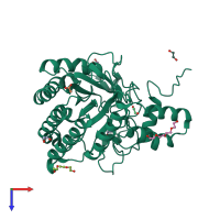
4i5r

X-ray diffraction
1.5Å resolution

Crystal structure of a fungal chimeric cellobiohydrolase Cel6A

Released:

Function and Biology Details

Reaction catalysed:
Hydrolysis of (1->4)-beta-D-glucosidic linkages in cellulose and cellotetraose, releasing cellobiose from the non-reducing ends of the chains
Biological process:
Cellular component:
  • not assigned

Structure analysis Details

Assembly composition:
monomeric (preferred)
PDBe Complex ID:
PDB-CPX-139870 (preferred)
Entry contents:
1 distinct polypeptide molecule
Macromolecule:
Exoglucanase 2; Exoglucanase-6A; Glucanase Chain: A
Molecule details ›
Chain: A
Length: 364 amino acids
Theoretical weight: 39.77 KDa
Source organisms: Expression system: Saccharomyces cerevisiae
UniProt:
  • Canonical: Q5G2D4 (Residues: 118-121, 339-435; Coverage: 22%)
  • Canonical: Q9C1S9 (Residues: 121-161; Coverage: 9%)
  • Canonical: P07987 (Residues: 158-331, 430-471; Coverage: 48%)
Gene names: avi2, cbh2, cel6A
Sequence domains: Glycosyl hydrolases family 6
Structure domains: 1, 4-beta cellobiohydrolase

Ligands and Environments

No modified residues

Experiments and Validation Details

Entry percentile scores
X-ray source: SSRL BEAMLINE BL12-2
Spacegroup: C2
Unit cell:
a: 158.61Å b: 45.262Å c: 58.602Å
α: 90° β: 95.21° γ: 90°
R-values:
R R work R free
0.156 0.154 0.181
Expression system: Saccharomyces cerevisiae